. 2013 Nov;195(22):5041-50.
doi: 10.1128/JB.00505-13. Epub 2013 Aug 30.

Regulation of host hemoglobin binding by the Staphylococcus aureus Clp proteolytic system

Affiliations

Regulation of host hemoglobin binding by the Staphylococcus aureus Clp proteolytic system

Allison J Farrand et al. J Bacteriol. 2013 Nov.

Abstract

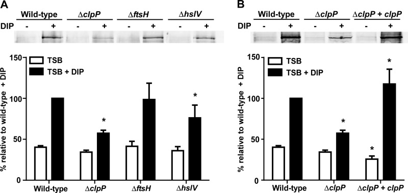
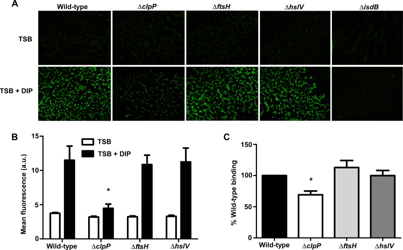
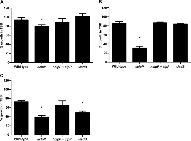
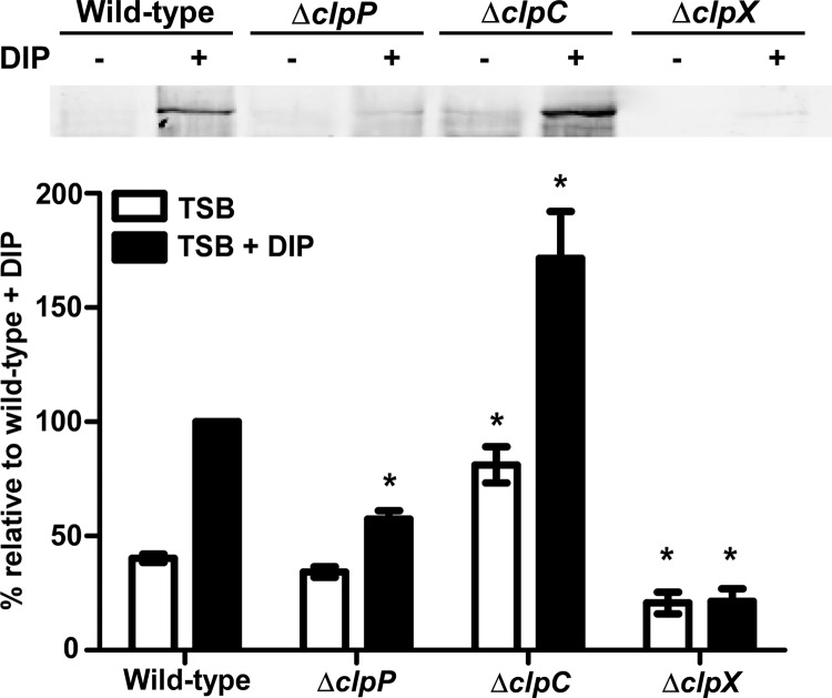
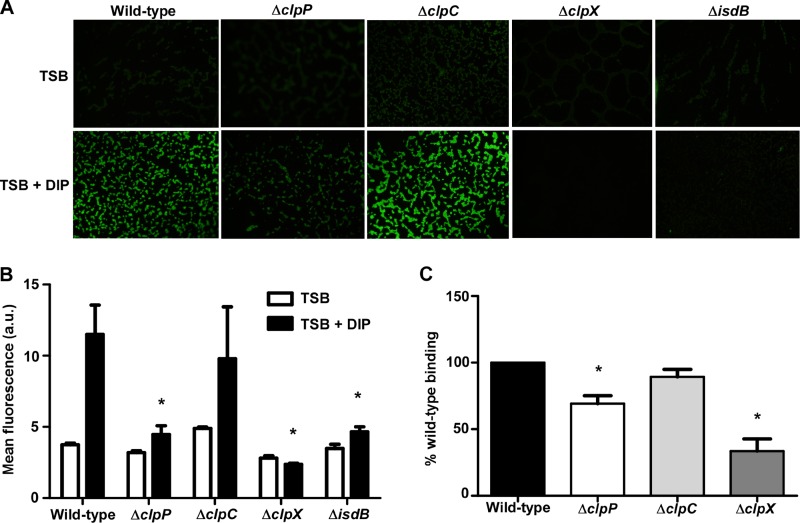
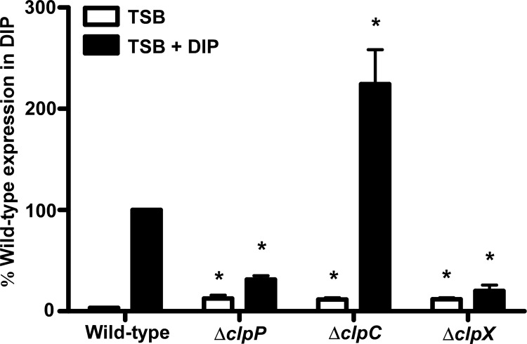
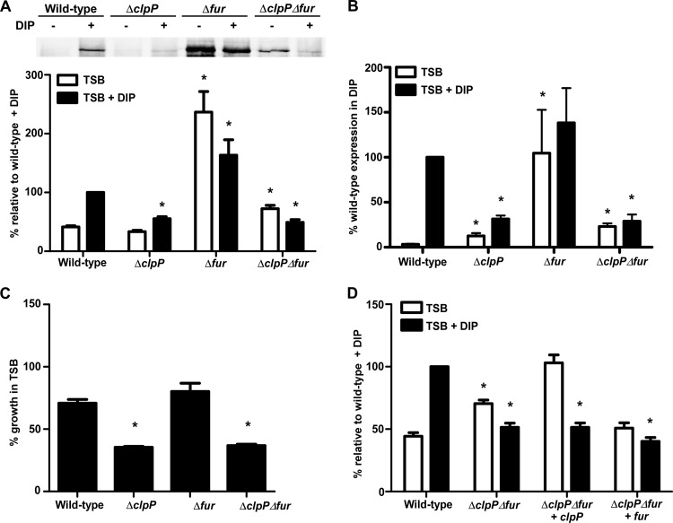
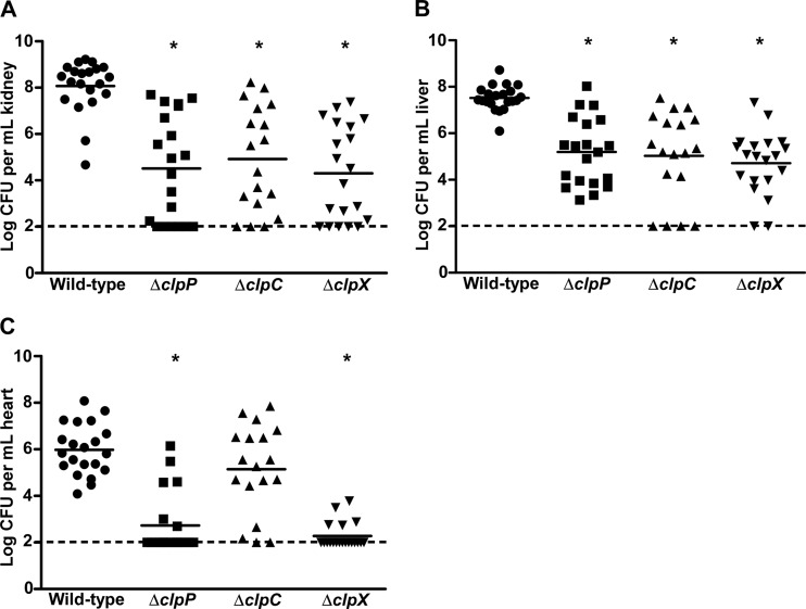
Protein turnover is a key process for bacterial survival mediated by intracellular proteases. Proteolytic degradation reduces the levels of unfolded and misfolded peptides that accumulate in the cell during stress conditions. Three intracellular proteases, ClpP, HslV, and FtsH, have been identified in the Gram-positive bacterium Staphylococcus aureus, a pathogen responsible for significant morbidity and mortality worldwide. Consistent with their crucial role in protein turnover, ClpP, HslV, and FtsH affect a number of cellular processes, including metabolism, stress responses, and virulence. The ClpP protease is believed to be the principal degradation machinery in S. aureus. This study sought to identify the effect of the Clp protease on the iron-regulated surface determinant (Isd) system, which extracts heme-iron from host hemoglobin during infection and is critical to S. aureus pathogenesis. Inactivation of components of the Clp protease alters abundance of several Isd proteins, including the hemoglobin receptor IsdB. Furthermore, the observed changes in IsdB abundance are the result of transcriptional regulation, since transcription of isdB is decreased by clpP or clpX inactivation. In contrast, inactivation of clpC enhances isdB transcription and protein abundance. Loss of clpP or clpX impairs host hemoglobin binding and utilization and results in severe virulence defects in a systemic mouse model of infection. These findings suggest that the Clp proteolytic system is important for regulating nutrient iron acquisition in S. aureus. The Clp protease and Isd complex are widely conserved in bacteria; therefore, these data reveal a novel Clp-dependent regulation pathway that may be present in other bacterial pathogens.


Figures

Fig 1
Fig 1
Disruption of the S. aureus clpP proteolytic subunit reduces abundance of the hemoglobin receptor IsdB. (A) IsdB protein levels analyzed by immunoblotting. Wild-type S. aureus or strains inactivated for clpP, ftsH, or hslV were grown in TSB or TSB supplemented with the iron chelator DIP (1 mM). Total protein in cell wall fractions was normalized and separated by 15% SDS-PAGE. Proteins were transferred to nitrocellulose and probed with an anti-IsdB antibody. (Top) Blots are representative of at least 4 independent experiments. (Bottom) Graphical representation of IsdB protein abundance in TSB (white bars) or TSB plus DIP (black bars) as assessed by densitometry analysis of immunoblots. (B) Restoration of IsdB protein levels through complementation of clpP, assessed by immunoblotting (top) and densitometry (bottom). Data represent 4 independent experiments. Error bars represent standard errors of the means (SEM). *, P <0.025 relative to the wild type under the respective conditions, as calculated by Student's t test.
Fig 2
Fig 2
Disruption of the S. aureus clpP proteolytic subunit impairs binding of host hemoglobin. (A) Surface exposure of IsdB as analyzed using immunofluorescence microscopy. Wild-type S. aureus or protease knockouts were grown in iron-rich (TSB) or iron-poor (TSB + DIP) medium, fixed on coverslips, and probed with anti-IsdB and a secondary antibody conjugated to Alexa Fluor 488. Images are representative of at least 2 experiments. (B) Graphical representation of IsdB immunofluorescence. a.u., arbitrary units. *, P < 0.001. (C) Hemoglobin capture by wild-type or mutant S. aureus strains was determined using a whole-cell cosedimentation assay. Iron-starved cells were incubated with human hemoglobin, and captured hemoglobin was removed by pelleting cells, treating them with SDS, and boiling the samples. Bound hemoglobin was separated by SDS-PAGE, silver stained, and analyzed by densitometry. Data represent at least 5 experiments. Error bars represent SEM. *, P < 0.0001 relative to the wild type, as calculated by Student's t test.
Fig 3
Fig 3
ClpP is required for hemoglobin-dependent heme acquisition. Wild-type S. aureus, knockouts of clpP or isdB, and the complemented clpP mutant were grown in iron-depleted medium and supplemented with iron sulfate (A), heme (B), or hemoglobin (C). Growth was determined by measuring the optical density at 600 nm, and data are expressed as percent growth compared to that of each strain in iron-rich medium at 24 h. Data shown are means and SEM for three separate experiments. *, P < 0.0004 relative to the wild type, as calculated by Student's t test.
Fig 4
Fig 4
Inactivation of the Clp ATPases drastically alters IsdB protein levels. (Top) IsdB protein levels were analyzed by Western blotting. Wild-type S. aureus or strains inactivated for clpP, clpC, or clpX were grown in iron-rich (TSB) or iron-poor (TSB + DIP) medium. Total protein in cell wall fractions was normalized and separated by 15% SDS-PAGE. Proteins were transferred to nitrocellulose and probed with antibody specific to IsdB. Blots are representative of at least 4 independent experiments. (Bottom) Graphical representation of IsdB protein abundance in TSB (white bars) or TSB plus DIP (black bars) as assessed by densitometry analysis of Western blots. Error bars represent SEM. *, P < 0.0002 relative to the wild type under the respective conditions, as calculated by Student's t test.
Fig 5
Fig 5
Inactivation of clpX ATPase significantly impairs host hemoglobin binding. (A) Surface exposure of IsdB as determined by immunofluorescence microscopy. Wild-type S. aureus or strains inactivated for components of the Clp proteolytic system were grown in iron-rich or iron-poor medium, fixed on coverslips, and probed with anti-IsdB and a secondary antibody conjugated to Alexa Fluor 488. Images are representative of at least 2 experiments. (B) Graphical representation of IsdB immunofluorescence. *, P < 0.003. (C) Hemoglobin capture of wild-type or mutant S. aureus strains was determined using a whole-cell cosedimentation assay. Iron-starved cells were incubated with human hemoglobin, and captured hemoglobin was removed by pelleting cells, treating them with SDS, and boiling the samples. Bound hemoglobin was separated by SDS-PAGE, and gels were silver stained and analyzed by densitometry. Data represent at least 5 experiments. Error bars represent SEM. *, P < 0.0001 relative to the wild type, as calculated by Student's t test.
Fig 6
Fig 6
Inactivation of clp genes alters isdB expression. Expression of isdB was measured using qRT-PCR. Wild-type S. aureus or strains inactivated for clpP, clpC, or clpX were grown in TSB (white bars) or TSB with 1 mM DIP (black bars) to late exponential phase. RNAs were isolated from samples and converted to cDNAs. Expression of isdB was analyzed using primers specific to the isdB coding sequence. Data are expressed as fold transcript levels relative to wild-type isdB expression in TSB, which was set at 1. The graph represents data from 3 individual experiments. Error bars represent SEM. *, P < 0.007 relative to the wild type under the respective conditions, as calculated by Student's t test.
Fig 7
Fig 7
Clp proteins influence IsdB independently of Fur. (A) (Top) IsdB protein levels were analyzed by immunoblotting. Wild-type S. aureus or strains inactivated for fur alone or in combination with the knockout of clpP were grown in TSB alone or in the presence of DIP (1 mM), and cell wall fractions were blotted for IsdB. Blots are representative of 4 independent experiments. (Bottom) Graphical representation of IsdB protein abundance in TSB (white bars) or TSB plus DIP (black bars) as determined by densitometry analysis of Western blots. (B) Quantitative RT-PCR analysis of isdB expression in fur mutants. Error bars represent SEM. *, P < 0.015 relative to the wild type under the respective conditions, as calculated by Student's t test. (C) Wild-type or mutant strains were assessed for growth with hemoglobin as the sole iron source. *, P < 0.001 relative to the wild type at 24 h. (D) IsdB protein levels in ΔclpP Δfur mutants complemented with either clpP or fur on a plasmid were determined by Western blotting.
Fig 8
Fig 8
Clp proteolytic system is required for staphylococcal pathogenesis in a systemic infection model. Seven-week-old female BALB/c mice were retro-orbitally infected with 107 CFU wild-type S. aureus Newman or strains inactivated for clpP, clpC, or clpX. Infection progressed for 96 h, at which time mice were sacrificed, organs harvested, and bacterial burdens determined by serial dilution and plating. Numbers of recovered CFU from infected kidneys (A), livers (B), and hearts (C) are shown. Graphs include data from two individual experiments with a total of 20 mice. The dashed lines denote the limit of detection. Error bars represent SEM. *, P < 0.0001 relative to the wild type, as calculated by Student's t test.

References

    1. Moran GJ, Krishnadasan A, Gorwitz RJ, Fosheim GE, McDougal LK, Carey RB, Talan DA. 2006. Methicillin-resistant S. aureus infections among patients in the emergency department. N. Engl. J. Med. 355:666–674 - PubMed
    1. Miro JM, Anguera I, Cabell CH, Chen AY, Stafford JA, Corey GR, Olaison L, Eykyn S, Hoen B, Abrutyn E, Raoult D, Bayer A, Fowler VG, International Collaboration on Endocarditis Merged Database Group 2005. Staphylococcus aureus native valve infective endocarditis: report of 566 episodes from the International Collaboration on Endocarditis Merged Database. Clin. Infect. Dis. 41:507–514 - PubMed
    1. Purcell K, Fergie J. 2005. Epidemic of community-acquired methicillin-resistant Staphylococcus aureus infections: a 14-year study at Driscoll Children's Hospital. Arch. Pediatr. Adolesc. Med. 159:980–985 - PubMed
    1. Herold BC, Immergluck LC, Maranan MC, Lauderdale DS, Gaskin RE, Boyle-Vavra S, Leitch CD, Daum RS. 1998. Community-acquired methicillin-resistant Staphylococcus aureus in children with no identified predisposing risk. JAMA 279:593–598 - PubMed
    1. Crosa J. 1997. Signal transduction and transcriptional and posttranscriptional control of iron-regulated genes in bacteria. Microbiol. Mol. Biol. Rev. 61:319–336 - PMC - PubMed

Publication types

MeSH terms