. 2013 Sep 19;501(7467):385-90.
doi: 10.1038/nature12521. Epub 2013 Sep 1.

Structural insight into the biogenesis of β-barrel membrane proteins

Affiliations

Structural insight into the biogenesis of β-barrel membrane proteins

Nicholas Noinaj et al. Nature. .

Abstract

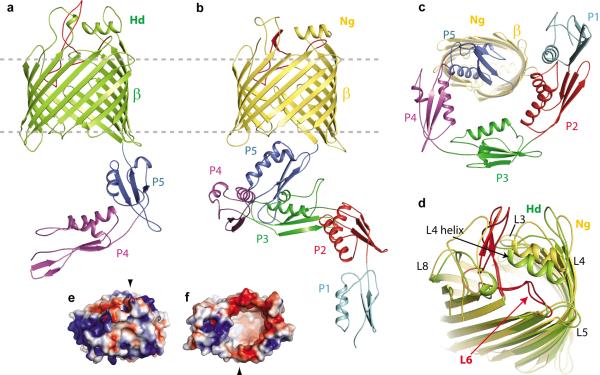
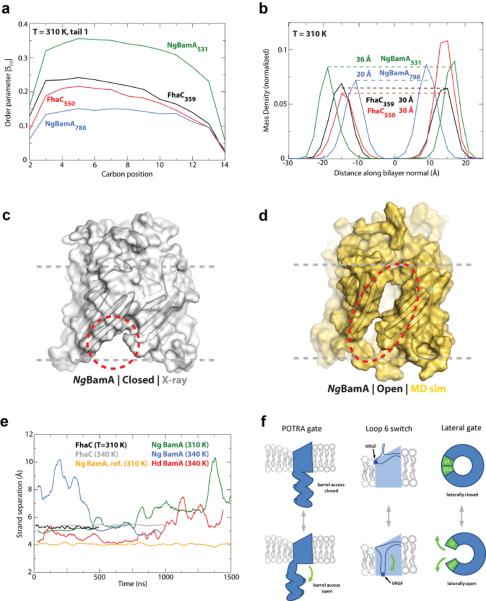
β-barrel membrane proteins are essential for nutrient import, signalling, motility and survival. In Gram-negative bacteria, the β-barrel assembly machinery (BAM) complex is responsible for the biogenesis of β-barrel membrane proteins, with homologous complexes found in mitochondria and chloroplasts. Here we describe the structure of BamA, the central and essential component of the BAM complex, from two species of bacteria: Neisseria gonorrhoeae and Haemophilus ducreyi. BamA consists of a large periplasmic domain attached to a 16-strand transmembrane β-barrel domain. Three structural features shed light on the mechanism by which BamA catalyses β-barrel assembly. First, the interior cavity is accessible in one BamA structure and conformationally closed in the other. Second, an exterior rim of the β-barrel has a distinctly narrowed hydrophobic surface, locally destabilizing the outer membrane. And third, the β-barrel can undergo lateral opening, suggesting a route from the interior cavity in BamA into the outer membrane.


Figures

Figure 1
Figure 1. The structure of BamA from the BAM complex
a. The HdBamAΔ3 crystal structure in cartoon representation showing the β-barrel (green) and POTRA domains 4 and 5 (purple and blue). b. The NgBamA crystal structure showing the β-barrel (gold) and POTRA domains 1–5 (cyan, red, green, purple and blue). c. A periplasmic (bottom) view of the NgBamA crystal structure. d. An alignment of the HdBamAΔ3 (green) and NgBamA (gold) crystal structures highlighting the structural conservation of the extracellular loops and secondary structural elements in loops 4 and 6. e. Electrostatic surface representation of HdBamAΔ3 viewed from the extracellular face and from the inside of the barrel from the periplasmic face (f). Black arrows indicate the locations of strand β-16.
Figure 2
Figure 2. HdBamA and NgBamA crystal structures reveal conformational changes
a. Alignment of HdBamAΔ3 (green) and NgBamA (gold) showing open and a closed conformations for the POTRA domains which may serve as a gating mechanism for regulating substrate access to the inside of the β-barrel. b. Compared to HdBamAΔ3 (green), strand β16 is disordered and tucked inside the β-barrel of NgBamA (gold). Arrowheads indicate the location of the C-terminal strand in Hd BamA (black) and NgBamA (red). Membrane view (c) and extracellular view (d) of an alignment of NgBamA and FhaC (gray, PDB code 2QDZ) illustrates conformational differences in the β-barrel and POTRA domains. In FhaC, the N-terminal α-helix (red) and loop 6 occlude the β-barrel preventing free diffusion across the outer membrane, however, in BamA this is accomplished by the extracellular loops that fold over the top of the barrel. Loop 6 (eL6) assumes different conformations between the two structures: e, membrane view; f zoomed view; g, periplasmic view. Unlike FhaC, NgBamA eL6 contains a β-hairpin (dashed circle) and the VRGF/Y motif is located ~18 Å from the periplasmic boundary. Unzipping of this β-hairpin may allow for an extended conformation similar to what is observed for eL6 of FhaC.
Figure 3
Figure 3. Mutational analysis of E.coli BamA
a. A homology model of EcBamA showing the conserved VRGF/Y and FQF motifs and putative R661 ionic interactions. b. Colony growth assays of cells reliant upon mutant EcBamA in the absence of arabinose: (1) WT EcBamA, (2) pRSF1 vector control, (3) R661A, (4) VRGF>A, (5) D740R, (6) E717,D740>A, and (7) ΔeL6 (residues 676–700). c. Growth curves of colonies isolated from +arabinose plates and then transferred to −arabinose rich medium. Error bars represent ± standard error of the mean. d. Western blots of EcBamA mutant expression levels (α-His) and DegP up-regulation when grown in −arabinose M63 minimal media. GroEL served as a loading control; numbers represent DegP fold-increases over WT. e. Heat modifiability of EcBamA mutants expressed in LB +arabinose, and of LamB after arabinose wash-out (at the 4 hour time point as in panel c where no growth was observed for the vector control, VRGF>A, or D740R). All His-tagged EcBamA mutants except VRGF>A and D740R showed evidence of folding and LamB trimer formation. f. α-His reactive cleavage products (*) following proteinase K proteolysis of whole cells indicated extracellular accessibility of all EcBamA mutants except VRGF>A. Dashes indicate full-length mutant BamA; arrowheads indicate a nonspecific α-His-HRP reactive protein. Maltose binding protein (MBP) was used to control for outer membrane integrity. All data shown is representative of at least three independent experiments.
Figure 4
Figure 4. BamA primes the membrane for OMP insertion
Molecular dynamics (MD) simulations investigated the stability of the BamA β-barrel. a. SCD values, a measure of lipid order, were decreased near NgBamA strand β16 (centered at residue 788) compared to the opposite side of the β-barrel (centered at residue 531). Minimal differences were observed for FhaC comparing analogous sites. b. MD analysis revealed that the β-barrel of NgBamA imparts a thinning of the membrane by 16 Å near strand β16 (centered at residue 788) when compared to the opposite side of the barrel (centered at residue 531), whereas, no difference was observed for FhaC. The MD simulations also indicated that the interaction between strands β1 and β16 is significantly destabilized, allowing the β-barrel to undergo a lateral opening (c and d, closed and open states, respectively). e. Quantification of the separation between strands β1 and β16 shows that both NgBamA (green) and HdBamA (red) structures have the propensity to open, unlike FhaC (black and grey). As reference, no change was observed between strands β13 and β14 for NgBamA (orange). f. Summary of the putative conformational switches of BamA based on structural and computational analysis. The first is the conformational gate of the POTRA domains (membrane view), the second is the conformational switch of loop 6 from a resting state (observed in our crystal structures) to a putative activated state as observed in FhaC, potentially representing the protease-sensitive state observed by Rigel et al., and the third is the lateral opening event (extracellular surface view).

Comment in

References

    1. Dalbey RE, Wang P, Kuhn A. Assembly of bacterial inner membrane proteins. Annu Rev Biochem. 2011;80:161–187. - PubMed
    1. White SH, von Heijne G. How translocons select transmembrane helices. Annu Rev Biophys. 2008;37:23–42. - PubMed
    1. du Plessis DJ, Nouwen N, Driessen AJ. The Sec translocase. Biochim Biophys Acta. 2011;1808:851–865. - PubMed
    1. Osborne AR, Rapoport TA, van den Berg B. Protein translocation by the Sec61/SecY channel. Annu Rev Cell Dev Biol. 2005;21:529–550. - PubMed
    1. Chacinska A, Koehler CM, Milenkovic D, Lithgow T, Pfanner N. Importing mitochondrial proteins: machineries and mechanisms. Cell. 2009;138:628–644. - PMC - PubMed

Publication types

MeSH terms

Substances