. 2013 Nov;9(11):677-84.
doi: 10.1038/nchembio.1335. Epub 2013 Sep 1.

Paralog-selective Hsp90 inhibitors define tumor-specific regulation of HER2

Affiliations

Paralog-selective Hsp90 inhibitors define tumor-specific regulation of HER2

Pallav D Patel et al. Nat Chem Biol. 2013 Nov.

Abstract

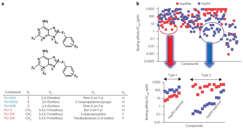
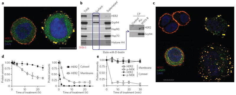
Although the Hsp90 chaperone family, comprised in humans of four paralogs, Hsp90α, Hsp90β, Grp94 and Trap-1, has important roles in malignancy, the contribution of each paralog to the cancer phenotype is poorly understood. This is in large part because reagents to study paralog-specific functions in cancer cells have been unavailable. Here we combine compound library screening with structural and computational analyses to identify purine-based chemical tools that are specific for Hsp90 paralogs. We show that Grp94 selectivity is due to the insertion of these compounds into a new allosteric pocket. We use these tools to demonstrate that cancer cells use individual Hsp90 paralogs to regulate a client protein in a tumor-specific manner and in response to proteome alterations. Finally, we provide new mechanistic evidence explaining why selective Grp94 inhibition is particularly efficacious in certain breast cancers.


Conflict of interest statement

Competing financial interests

The authors declare competing financial interests: details accompany the online version of the paper.

Figures

Figure 1
Figure 1. Library screening identifies paralog specific chemical spaces
(a) General structure of the purine-scaffold chemical library. Screening identified the Grp94-selective compounds PU-H54, PU-WS13 and PU-H39 (5) and the Hsp90-selective compounds PU-11 (6), PU-29F and PU-20F (7). All but PU-H54 have X3 attached at N9. (b) Top, graphical representation of screening data from the paralog-specific fluorescence polarization assay. Over 130 purine-scaffold compounds were screened. Each x-axis entry represents a different compound. Compounds were tested in triplicate. Bottom, 21 paralog-selective purine-scaffold compounds were identified. Several Grp94-selective compounds (Type 2) had greater than 100-fold preference for Grp94 over Hsp90α and Hsp90β and a 10- to 100-fold preference over Trap-1 (Supplementary Fig. 1b,c). In the Hsp90α-selective subtype (Type 1), we found ligands that showed a 5- to 15-fold and a 5- to 35-fold selectivity over Grp94 and Trap-1, respectively. Unexpectedly, in spite of the high homology between Hsp90α and Hsp90β, we also identified compounds with three- to fivefold selectivity for the Hsp90α paralog (Supplementary Fig. 2b,c). Detailed structures and binding data are presented in Supplementary Figures 1 and 2.
Figure 2
Figure 2. Structural and computational analyses define differences in Hsp90 paralog pockets and paralog-selective chemical spaces
(a) Overlay of Hsp90α- and Grp94-bound PU-H54 reveals an 80° torsional rotation about the sulfanyl linker (highlighted in red) when inserted into the Grp94-specific channel. (b) Interactions of PU-H54 bound to Grp94 showing the increased hydrophobic stabilization of the 8-aryl group (X2-Ar) when bound into Site 2. (c) Surface area (left) and conformation (right) of paralog-selective chemical spaces as generated by Macromodel/Molecular Surface. Figures were created by superimposing the favored docking pose of all of the Hsp90α (top, Type 1) and Grp94 (bottom, Type 2) inhibitors described in Supplementary Figures 1 and 2.
Figure 3
Figure 3. HER2 is sensitive to Hsp90 paralog inhibition in a tumor-specific manner
(a) SKBr3 (left graph, high HER2) and MCF7 (right graph, low HER2) cells were treated for 24 h with vehicle (DMSO), the Grp94-selective inhibitors PU-WS13 and PU-H39, the Hsp90α- and Hsp90β-selective inhibitors PU-29F and PU-20F, the pan-Hsp90 inhibitor PU-H71 or for 72 h with siRNAs specific for Grp94 or Hsp90α and Hsp90β. Amounts of HER2 are plotted for each experimental condition. Associated representative western blots (WBs) are shown in Supplementary Figure 7a,b. Control siRNA, scramble construct. Data are presented as mean ± s.d. (n = 3). (b) Representative western blot of HER2 complexes in MCF7 extracts isolated by precipitation with an antibody to HER2 (anti-HER2) or a nonspecific IgG. IP, immunoprecipitation; rIgG, rabbit IgG, an isotype control. (c) MCF7 cells treated for the indicated times with the Hsp90α- and Hsp90β-selective PU-11 (40 μM) or the Grp94-selective PU-WS13 (15 μM). Protein amounts in membrane and cytosolic fractions were plotted against the time of treatment. Data are presented as mean ± s.e.m. (n = 3). Associated representative western blots are shown in Supplementary Figure 7f. Full gels of key experiments are shown in Supplementary Note 1.
Figure 4
Figure 4. Grp94 and Hsp90α and Hsp90β regulate distinct HER2 functions in HER2-overexpressing cancer cells
(a) Fluorescence microscopy image of SKBr3 cells treated for 4 h with DMSO (left) or PU-WS13 (right, 15 μM) and then stained with the indicated markers upon fixation and permeabilization. Scale bar, 10 μm. (b) Representative blot of surface-exposed proteins chemically labeled with biotin and purified using streptavidin columns. Histone H4 was blotted to control for membrane impermeability. Proteins eluted from the streptavidin column were affinity purified and analyzed by western blotting as indicated. Total, total cell extracts; supernatant; nonsurface proteins; PU-WS13B, PW-WS13-biotin immobilized on streptavidin beads; CP, chemical precipitation. (c) Same as in a for cells treated with DMSO (top left), PU-WS13 (top right, 15 μM) PU-29F (bottom left, 20 μM) or PU-H71 (bottom right, 1 μM). Scale bar, 10 μm. Additional stains are shown in Supplementary Figure 8d,e. (d,e) SKBr3 cells were treated for the indicated times with 20 μM of the Grp94-selective compound PU-WS13 (d, left, and e) or the Hsp90α- and Hsp90β-selective PU-29F (d, right). Proteins in membrane and cytosolic fractions were plotted against the time of treatment. Data are presented as mean ± s.e.m. (n = 3). Associated representative western blots are shown in Supplementary Figure 8g,h. Full gels of key experiments are shown in Supplementary Note 1.
Figure 5
Figure 5. Schematic representation summarizing the tumor-specific regulation of HER2 by the Hsp90 paralogs
All epithelial cells contain two copies of the HER2-encoding gene and express small amounts of the HER2 receptor on the cell surface. During oncogenic transformation, the number of gene copies per cell may increase, as in the SKBr3 cell line, leading to an increase in mRNA transcription and a 100- to 1,000-fold increase in the number of HER2 receptors on the cell surface. Our data suggest a tumor-specific involvement of the Hsp90 paralogs in the chaperoning of HER2. The Hsp90α paralog is sufficient for HER2 trafficking and function in cells with low HER2 expression such as MCF7. In contrast, in cells with excessive amounts of HER2, such as in SKBr3, all three paralogs have an important role, with Hsp90α and Hsp90β regulating cytosolic HER2 and Grp94 regulating plasma membrane HER2. Because HER2 is the major oncogene in these cells, its dependence on Grp94 renders cells addicted to Grp94, proposing Grp94 as a new target in such cancers.
Figure 6
Figure 6. Grp94 inhibition alone is sufficient to induce apoptosis in and reduce the viability of HER2 overexpressing breast cancer cells
(a,b) Viability of breast cancer cells in which Grp94 was inhibited with PU-WS13 (a) or knocked down by means of siRNA (b). Cell viability was assessed using an assay that quantifies ATP. A representative western blot indicating protein changes in SKBr3 cells upon Grp94 knockdown is also shown in b. (c) Representative western blots of cancer cells treated for 24 h with PU-WS13 (0.5 μM, 2.5 μM and 12.5 μM) or vehicle. Full gels of key experiments are shown in Supplementary Note 1.

References

    1. Workman P, Burrows F, Neckers L, Rosen N. Drugging the cancer chaperone Hsp90: combinatorial therapeutic exploitation of oncogene addiction and tumor stress. Ann NY Acad Sci. 2007;1113:202–216. - PubMed
    1. Sreedhar AS, Kalmar E, Csermely P, Shen YF. Hsp90 isoforms: functions, expression and clinical importance. FEBS Lett. 2004;562:11–15. - PubMed
    1. Johnson JL. Evolution and function of diverse Hsp90 homologs and cochaperone proteins. Biochim Biophys Acta. 2012;1823:607–613. - PubMed
    1. Chène P. ATPases as drug targets: learning from their structure. Nat Rev Drug Discov. 2002;1:665–673. - PubMed
    1. Pearl LH, Prodromou C, Workman P. The Hsp90 molecular chaperone: an open and shut case for treatment. Biochem J. 2008;410:439–453. - PubMed

Publication types

MeSH terms