. 2014 Oct;48(19):1414-20.
doi: 10.1136/bjsports-2013-092438. Epub 2013 Aug 30.

Substance P reduces TNF-α-induced apoptosis in human tenocytes through NK-1 receptor stimulation

Affiliations
Free PMC article

Substance P reduces TNF-α-induced apoptosis in human tenocytes through NK-1 receptor stimulation

Ludvig J Backman et al. Br J Sports Med. 2014 Oct.
Free PMC article

Abstract

Background: It has been hypothesised that an upregulation of the neuropeptide substance P (SP) and its preferred receptor, the neurokinin-1 receptor (NK-1 R), is a causative factor in inducing tenocyte hypercellularity, a characteristic of tendinosis, through both proliferative and antiapoptotic stimuli. We have demonstrated earlier that SP stimulates proliferation of human tenocytes in culture.

Aim: The aim of this study was to investigate whether SP can mediate an antiapoptotic effect in tumour necrosis factor-α (TNF-α)-induced apoptosis of human tenocytes in vitro.

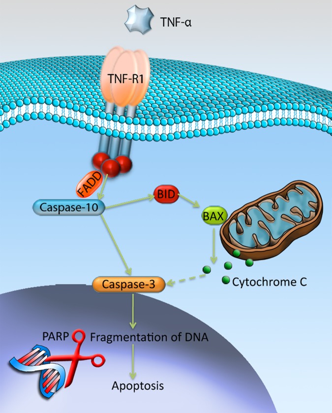
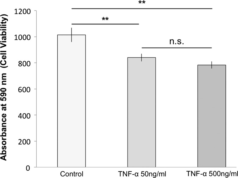
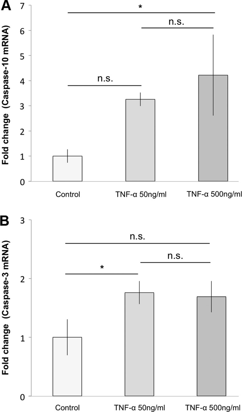
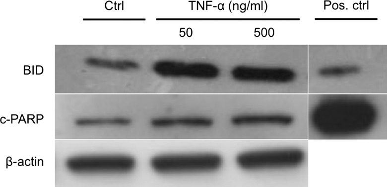
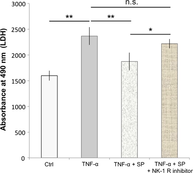
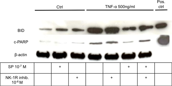
Results: A majority (approximately 75%) of tenocytes in culture were immunopositive for TNF Receptor-1 and TNF Receptor-2. Exposure of the cells to TNF-α significantly decreased cell viability, as shown with crystal violet staining. TNF-α furthermore significantly increased the amount of caspase-10 and caspase-3 mRNA, as well as both BID and cleaved-poly ADP ribosome polymerase (c-PARP) protein. Incubation of SP together with TNF-α resulted in a decreased amount of BID and c-PARP, and in a reduced lactate dehydrogenase release, as compared to incubation with TNF-α alone. The SP effect was blocked with a NK-1 R inhibitor.

Discussion: This study shows that SP, through stimulation of the NK-1 R, has the ability to reduce TNF-α-induced apoptosis of human tenocytes. Considering that SP has previously been shown to stimulate tenocyte proliferation, the study confirms SP as a potent regulator of cell-turnover in tendon tissue, capable of stimulating hypercellularity through different mechanisms. This gives further support for the theory that the upregulated amount of SP seen in tendinosis could contribute to hypercellularity.

Keywords: Achilles Tendon; Tendons.


Figures

Figure 1
Figure 1
Mechanisms of tumour necrosis factor-α (TNF-α)-induced apoptosis through the TNF-R1. Binding of TNF-α to TNF-R1 results in activation of caspase-10 through recruitment of the Fas-Associated protein with Death Domain (FADD). Activated caspase-10 has the ability to activate caspase-3, either directly by cleavage, or indirectly by activating the Bcl-2 family protein BID that through release of cytochrome-c will activate caspase-3. Ultimately, activated caspase-3 will result in fragmentation of DNA, that is, apoptosis, with the involvement of cleaved-poly ADP ribosome polymerase (PARP). Illustration by Gustav Andersson (copyright with artist).
Figure 2
Figure 2
Tumour necrosis factor-α (TNF-α) receptors in cultured human tenocytes. Immunocytochemistry to confirm the presence of TNF-R1 (A) and TNF-R2 (B) in cultured human tenocytes (green; FITC). Cells are counterstained with DAPI (blue) to mark the nuclei (×600).
Figure 3
Figure 3
Reduced cell viability after tumour necrosis factor-α (TNF-α) incubation. A significant decrease in cell viability of cultured human tenocytes was seen in cells treated with both 50 ng/mL of TNF-α and 500 ng/mL TNF-α, as compared to the control after 72 h of exposure (**p<0.01). The difference in cell viability seen between the treatments with different concentrations of TNF-α was not statistically significant (n.s.; p=0.07). Cell viability was measured with crystal violet staining (A590). Error bars indicate SD.
Figure 4
Figure 4
Effects of tumour necrosis factor-α (TNF-α) on the apoptotic signalling pathway at mRNA level. Subjecting cultured human tenocytes to TNF-α for 7 h significantly increased main parameters of the apoptotic pathway. (A) 500 ng/mL of TNF-α significantly increased caspase-10 mRNA as compared to the control (4.2-fold change; *p<0.05). 50 ng/mL also showed an increase compared to control, although this was not statistically significant (n.s) (p=0.08). (B) In total, 50 ng/mL of TNF-α significantly increased caspase-3 mRNA in comparison to the control (1.8-fold change; *p<0.05). The increase seen for cells treated with 500 ng/mL of TNF-α was not statistically significant (n.s.), although close to significant (p=0.05). Error bars indicate the SD.
Figure 5
Figure 5
Effects of tumour necrosis factor-α (TNF-α) on the apoptotic signalling pathway at protein level. Twelve hours of exposure to TNF-α resulted in an increase of both BID and cleaved-PARP (c-PARP) in cultured human tenocytes. A more obvious increase in comparison to the control (Ctrl) was seen for BID, as compared to c-PARP, after treatment with TNF-α. β-Actin was used as a loading control. Jurkat apoptosis cell lysate was used as a positive control (Pos. ctrl).
Figure 6
Figure 6
Substance P (SP) reduces TNF-α-induced cell death. Human cultured tenocytes treated with 500 ng/mL of TNF-α for 48 h showed a significant increase in the amount of lactate dehydrogenase (LDH)-release compared to the control (Ctrl). Pretreatment with SP at concentrations of 10−7 M significantly reduced the TNF-α-induced LDH-release, as compared to TNF-α alone. However, when the neurokinin-1 receptor (NK-1 R) inhibitor (at concentrations of 10−6 M) was added to the SP and TNF-α treatments of the cells, the LDH release was not significantly (n.s.) different from that after incubation with TNF-α alone, but significantly increased as compared to incubation with TNF-α together with SP. Thus, the SP effect of reducing the TNF-α-induced LDH-release, that is, cell death, was effectively inhibited by blocking the NK-1 R, showing that the SP effect is mediated through this receptor. Error bars indicate SD (*p<0.05; **p<0.01).
Figure 7
Figure 7
Substance P (SP) reduces the tumour necrosis factor-α (TNF-α)-induced apoptosis in human tenocytes. The increase in expression of BID and cleaved-PARP seen in cultured human tenocytes after incubation with TNF-α as compared to control (Ctrl), was clearly reduced when cells were pretreated with SP. Blocking of the SP receptor with an NK-1 R inhibitor (NK-1 R inhib.) reduced this effect of SP. β-actin was used as a loading control and jurkat apoptosis cell lysate was used as a positive control (Pos. ctrl; 12 h of incubations).

References

    1. Danielson P. Reviving the biochemical hypothesis for tendinopathy: new findings suggest the involvement of locally produced signal substances. Br J Sports Med 2009;43:265–8 - PubMed
    1. Khan KM, Cook JL, Maffulli N, et al. Where is the pain coming from in tendinopathy? It may be biochemical, not only structural, in origin. Br J Sports Med 2000;34:81–3 - PMC - PubMed
    1. Bjur D, Danielson P, Alfredson H, et al. Immunohistochemical and in situ hybridization observations favor a local catecholamine production in the human Achilles tendon. Histol Histopathol 2008;23:197–208 - PubMed
    1. Danielson P, Alfredson H, Forsgren S. Immunohistochemical and histochemical findings favoring the occurrence of autocrine/paracrine as well as nerve-related cholinergic effects in chronic painful patellar tendon tendinosis. Microsc Res Tech 2006;69:808–19 - PubMed
    1. Danielson P, Alfredson H, Forsgren S. Studies on the importance of sympathetic innervation, adrenergic receptors, and a possible local catecholamine production in the development of patellar tendinopathy (tendinosis) in man. Microsc Res Tech 2007;70:310–24 - PubMed

Publication types

MeSH terms

Substances