Review
. 2013 Oct;42(10):731-55.
doi: 10.1007/s00249-013-0925-x. Epub 2013 Sep 1.

Helical membrane protein conformations and their environment

Affiliations
Review

Helical membrane protein conformations and their environment

Timothy A Cross et al. Eur Biophys J. 2013 Oct.

Abstract

Evidence that membrane proteins respond conformationally and functionally to their environment is growing. Structural models, by necessity, have been characterized in preparations where the protein has been removed from its native environment. Different structural methods have used various membrane mimetics that have recently included lipid bilayers as a more native-like environment. Structural tools applied to lipid bilayer-embedded integral proteins are informing us about important generic characteristics of how membrane proteins respond to the lipid environment as compared with their response to other nonlipid environments. Here, we review the current status of the field, with specific reference to observations of some well-studied α-helical membrane proteins, as a starting point to aid the development of possible generic principles for model refinement.


Figures

Fig. 1
Fig. 1
Membrane proteins may have significant extra-membranous portions that behave much like soluble proteins, and can receive signals from the extracellular environment and transmit them to the intracellular side through a membrane-embedded, close-packed α-helical, domain, as with an ionotropic glutamate receptor, [PDB 3KG2] (Sobolevsky et al. 2009). This review focuses on the native-like structure of the transmembrane domain of helical membrane proteins.
Fig. 2
Fig. 2
Computed dielectric constant as a function of distance from bilayer center (Z position) for a POPC bilayer. The average fatty acyl carbonyl position is about Z=16Å. The choline groups can extend to about Z=27Å. The vertical lines represent the error bars. Reproduced with permission from (Nymeyer and Zhou 2008).
Fig. 3
Fig. 3
Conformational exchange for dimeric gramicidin A in isotropic solvents is enhanced by more than a 1000 fold by the addition of water (a protic solvent) that can facilitate hydrogen bond exchange. The conformational exchange is observed by GCOSY spectra of various double helical gramicidin A conformations in dioxane (a non-protic solvent). The red resonances highlight the stable state in this environment and its corresponding conformation is highlighted in red. a-c). a, d-f) In the presence of approximately 1% water in dioxane the conversion occurs in less than 1 h. This suggests that conformational interconversion in native membranes may be hindered by the sparsity of water in the membrane interstices. Reproduced with permission from (Xu and Cross 1999).
Fig. 4
Fig. 4
Lipid bilayers exhibit a large range of dynamics exemplified by normalized order parameters. The profiles for different bilayers and the variations of the molecular order parameter, Smol, with the segment position. ○, 1,2-dipalmitoyl-sn-glycero-3-phosphocholine. △, 1-palmitoyl-2-oleoyl-sn-glycero-3-phosphocholine. □, 1,2-dipalmitoyl-sn-glycero-3-phosphoserine. ×, Acholeplasma laidlawii (Stockton et al 1977). Reproduced with permission from (Seelig and Browning 1978).
Fig. 5
Fig. 5
Key amino acids have preferential locations in membrane protein structures with respect to the lipid bilayer environment. For example, tyrosine and tryptophan sidechains interacting with the lipid interfacial regions from a porin (a) and gramicidin A (b). Reproduced with permission from Killian and von Heijne (2000).
Fig. 6
Fig. 6
a) Viral budding of Influenza from a raft-like domain (yellow region in (d)). Hemagglutinin (HA) and Neuraminidase (NA) are highly soluble in the raft-like domain and have limited solubility in the pink liquid crystalline domain of the membrane; b) Elongation of the budding virus; c) membrane scission facilitated by M2 (blue) that has low solubility in the raft-like domain and functions in liquid crystalline domains; d) M2 (blue dots) is thought to associate with the raft/non-raft border, potentially by its affinity for cholesterol. It is therefore clustered in the non-raft region (pink) at the neck of the viral bud (c) where it induces curvature in the liquid crystalline environment via its amphipathic helices on the inner surface of the cellular membrane. Reproduced with permission from Rossman and Lamb (Rossman and Lamb 2011).
Fig. 7
Fig. 7
Bilayer and non-bilayer lattices in membrane protein crystals. a) Despite the bilayer lattice for the Influenza A M2 protein (3BKD) significant electrostatic interactions (space-filling Arg and Glu residues) between antiparallel tetramers appear to distort the tetramer helices (Stouffer et al. 2008). b) The non-bilayer lattice of estrone sulfatase (1P49) showing three monomers with nearly orthogonal TM helices and significant inter-monomer interactions (Hernandez-Guzman et al. 2003). c) The energy coupling factor-type riboflavin transporter (3P5N) also forms a lattice in which the TM domain of one protein is rotated by ~90° with respect to its neighbor and inter-protein interactions include helices that pack together via an Ala motif (Zhang et al. 2010).
Fig. 8
Fig. 8
A few solution NMR structures of α-helical membrane proteins. a-c) Space filling depiction of the polar residues. a) Histidine kinase receptor, ArcB (2KSD) displaying hydrophilic residues facing the hydrophobic environment and outwardly curved helices, potentially following the inner surface of the micelle. b) The dimer of BCL2/Adenovirus E1B interacting protein 3 (2KA2) shows a pair of interhelical hydrogen bonds between His and Ser residues. c) The tetramer of the drug resistant V27A M2 protein (2KWX) shows His and Trp residues buried near the pseudo four-fold symmetry axis. Ser residues are near the would be membrane interfacial region. d) The trimeric structure of diacyl glycerol kinase (2KDC) displaying outward curved helices and no apparent use of the Ala motifs (Ala Ca and Cb shown in space-filling view) for helix-helix packing.
Fig. 9
Fig. 9
Hydrophobic match of the membrane environment with the protein TM domain. a) The histidine kinase receptor, KdpD (2KSF) displays a short helix as well as numerous hydrophilic residues on the external surface of the helical bundle leading to considerable H/D exchange (white space filling amide protons indicate sites that H/D exchange). b-d) Influenza A M2 protein structures. b) The TM domain characterized in lipid bilayers with amantadine bound in the pore (2KQT) displaying helical lengths that span the hydrophobic region of the membrane. c) The conductance domain of M2 (residues 22-62; 2L0J) displaying very similar helical tilt angles to that of 2KQT. d) The M2 conductance domain in DHPC micelles (2RLF) displaying helices with a small tilt angle and drug (green space filling) on the external surface of the protein.
Fig. 10
Fig. 10
Native-like bound lipids and detergents in the crystal lattice shown with space filling atoms. a) In the ligand gated ion channel (PDB: 3EAM) a large number of diacyl lipids diffract in an annulus around the protein. b) For the voltage gated Na+ channel (3RVY) a monoacyl detergent is bound per monomer in a crevice at the monomer junctions, thought to be a lipid binding site.
Fig. 11
Fig. 11
Protein-protein crystal contacts can lead to structural perturbations. a) The acid sensing ion channel (ASIC; 2QTS) has a three-fold pseudo-symmetric water soluble domain and a distorted asymmetric TM domain. The contacts between monomers are boxed and highlighted in (c). b) 5-lipoxygenase (2Q7M) is also a trimer and one of the four helices forms crystal contacts with a neighboring trimer leading to what appears to be a shift in the TM helix of nearly 10Å out of the hydrophobic environment and for a hydrophilic interhelical loop to penetrate nearly 10Å into the membrane.
Fig. 12
Fig. 12
Thin hydrophobic environments can lead to excessively tilted helices and even disrupted helical structures. a) The P2X4 structure (3I5D) shows kinked and highly tilted helices resulting in a hydrophobic dimension that is less than 20Å thick and fenestrations into the pore from the fatty acyl environment. b) The site 2 protease (3B4R) displays TM helices that are interrupted by short β-strand segments that expose polar backbone sites to the lipid interstices.
Fig. 13
Fig. 13
Weak hydrophobic environments are suggested by the presence of water and polar groups that appear in the hydrophobic domain of the membrane. The charged residues are shown as spheres. a) The metal chelate transporter (2NQ2) has a large number of water molecules (additional red spheres) crystallized in the vicinity of the hydrophobic helices in a structure that otherwise appears to have a native-like TM domain. b) The maltose transporter (3FH6) has a pair of incomplete helices that do not span the membrane – in a native environment these helices would not be disordered and would span the hydrophobic dimension. c&d) The disulfide bond forming protein DsbB has been characterized by solution NMR (c: 2K73) and XRD (d: 2ZUQ) showing the exposure of hydrophilic residues to the would be bilayer interior.
Fig. 14
Fig. 14
Potential Effects of the Lateral Pressure Profile. The atoms of the charged residues are displayed as spheres. a&b) The Zinc transporter, YiiP (2QFI) has two 6 helix bundles splayed when viewed orthogonal to the plane containing the bundles (a) such that lipids could diffuse into the structure. b) When viewed at an oblique angle the cavities within the bundles are substantial, especially in comparison to (c) the well packed helices of the pyrophosphatase (4A01) where again the cavities in the structure are displayed, but are much smaller.
Fig. 15
Fig. 15
Detergent molecules embedded in the protein structures. Colored spheres represent the detergent molecules. a) The M2 proton channel (3BKD) with two octylglucoside and a polyethylene glycol in the pore of the structure and between the tetrameric helices. b) The acid sensing ion channel (2QTS) that displays an asymmetric trimeric TM domain has three detergent molecules embedded in the structure, one of which is on the trimeric axis. c) The sodium calcium exchanger (3V5U) has several detergent molecules embedded within the structure.

References

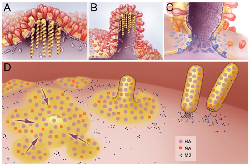
    1. Acharya R, Carnevale V, Fiorin G, Levine BG, Polishchuk AL, Balannik V, Samish I, Lamb RA, Pinto LH, DeGrado WF, Klein ML. Structure and mechanism of proton transport through the transmembrane tetrameric M2 protein bundle of the influenza A virus. Proceedings of the National Academy of Sciences of the United States of America. 2010;107(34):15075–15080. doi: 10.1073/pnas.1007071107. - DOI - PMC - PubMed
    1. Adams MN, Christensen ME, He Y, Waterhouse NJ, Hooper JD. The role of palmitoylation in signalling, cellular trafficking and plasma membrane localization of protease-activated receptor-2. PloS one. 2011;6(11):e28018. doi: 10.1371/journal.pone.0028018. - DOI - PMC - PubMed
    1. Agre P. The aquaporins: Blueprints tor cellular plumbing systems. Faseb J. 1999;13(7):A1520–A1520. - PubMed
    1. Andreas LB, Eddy MT, Pielak RM, Chou J, Griffin RG. Magic angle spinning NMR investigation of influenza A M2(18-60): support for an allosteric mechanism of inhibition. Journal of the American Chemical Society. 2010;132(32):10958–10960. doi: 10.1021/ja101537p. - DOI - PMC - PubMed
    1. Anfinsen CB. Principles that govern the folding of protein chains. Science. 1973;181(96):223–230. - PubMed

Publication types

MeSH terms