

A quantitative study of α-synuclein pathology in fifteen cases of dementia associated with Parkinson disease
- PMID: 23996276
- PMCID: PMC4041534
- DOI: 10.1007/s00702-013-1084-z
A quantitative study of α-synuclein pathology in fifteen cases of dementia associated with Parkinson disease
Abstract
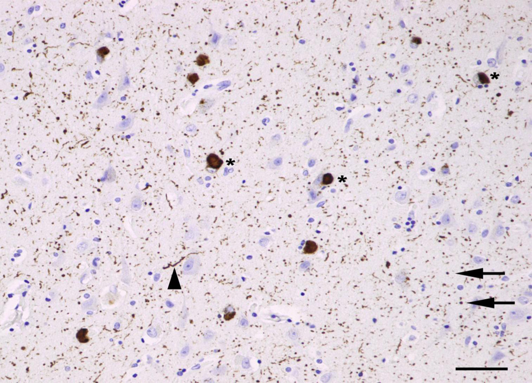
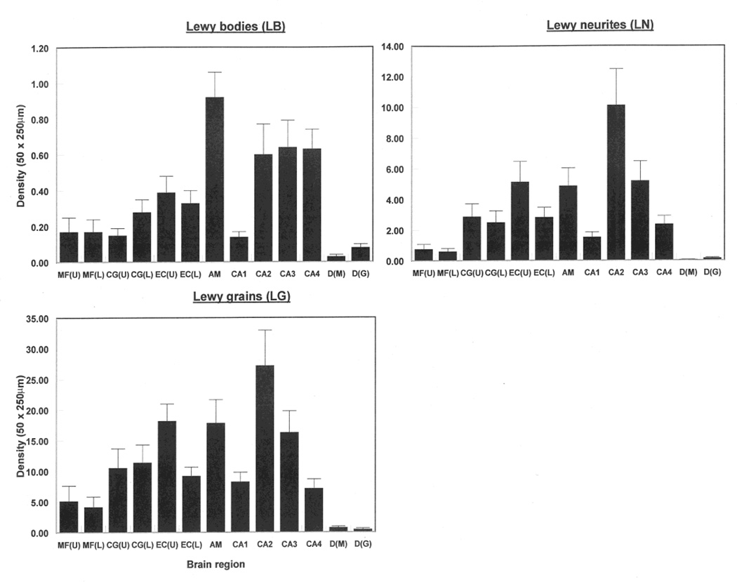
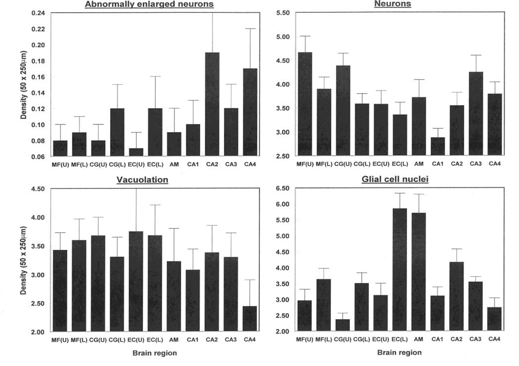
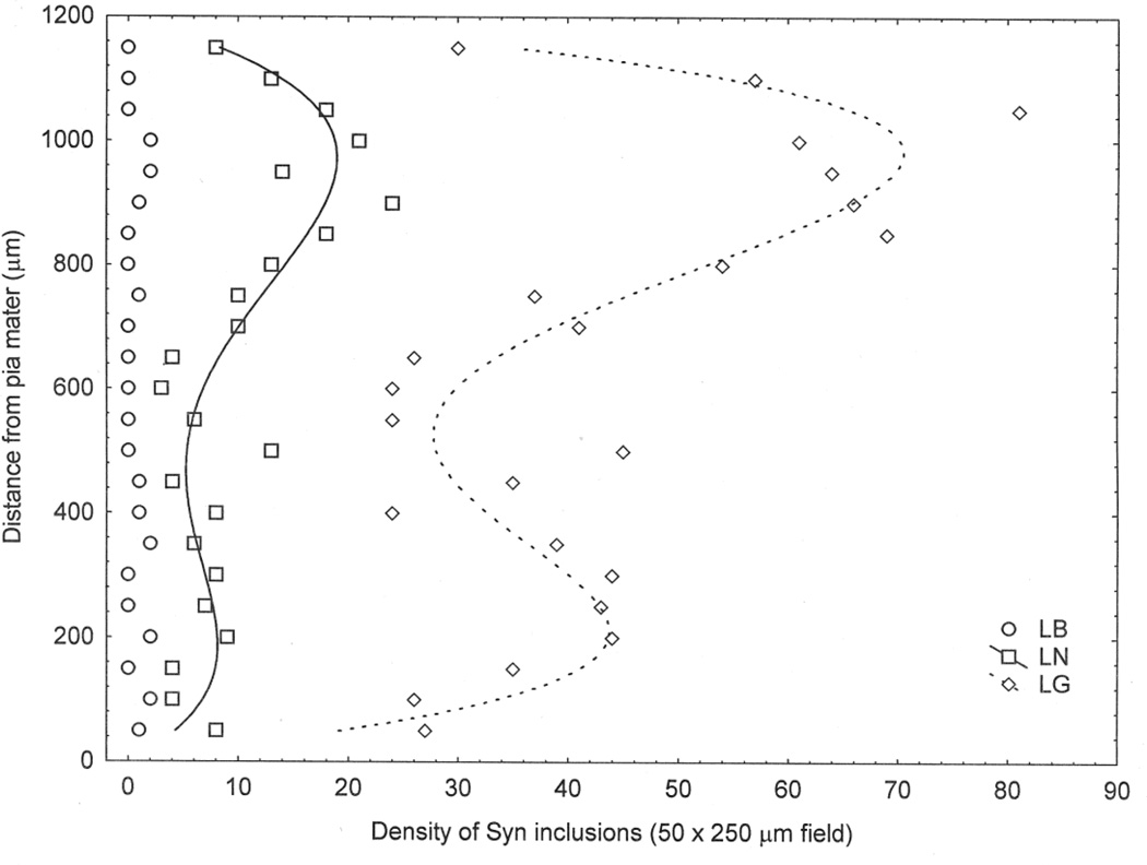
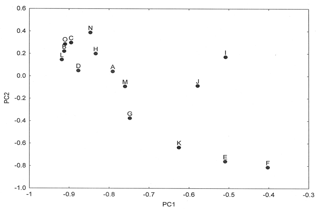
The α-synuclein-immunoreactive pathology of dementia associated with Parkinson disease (DPD) comprises Lewy bodies (LB), Lewy neurites (LN), and Lewy grains (LG). The densities of LB, LN, LG together with vacuoles, neurons, abnormally enlarged neurons (EN), and glial cell nuclei were measured in fifteen cases of DPD. Densities of LN and LG were up to 19 and 70 times those of LB, respectively, depending on region. Densities were significantly greater in amygdala, entorhinal cortex (EC), and sectors CA2/CA3 of the hippocampus, whereas middle frontal gyrus, sector CA1, and dentate gyrus were least affected. Low densities of vacuoles and EN were recorded in most regions. There were differences in the numerical density of neurons between regions, but no statistical difference between patients and controls. In the cortex, the density of LB and vacuoles was similar in upper and lower laminae, while the densities of LN and LG were greater in upper cortex. The densities of LB, LN, and LG were positively correlated. Principal components analysis suggested that DPD cases were heterogeneous with pathology primarily affecting either hippocampus or cortex. The data suggest in DPD: (1) ratio of LN and LG to LB varies between regions, (2) low densities of vacuoles and EN are present in most brain regions, (3) degeneration occurs across cortical laminae, upper laminae being particularly affected, (4) LB, LN and LG may represent degeneration of the same neurons, and (5) disease heterogeneity may result from variation in anatomical pathway affected by cell-to-cell transfer of α-synuclein.
Figures
References
-
- Antal A, Bandini P, Keri S, Bodis-Wollner I. Visuo-cognitive dysfunctions in Parkinson disease. Clin Neurosci. 1998;5:147–152. - PubMed
-
- Armstrong RA. Correlations between the morphology of diffuse and primitive β-amyloid (Aβ) deposits and the frequency of associated cells in Down’s syndrome. Neuropath Appl Neurobiol. 1996;22:527–530. - PubMed
-
- Armstrong RA. Quantifying the pathology of neurodegenerative disorders: quantitative measurements, sampling strategies and data analysis. Histopathol. 2003;42:521–529. - PubMed
-
- Armstrong RA. Visual signs and symptoms of Parkinson disease. Clin Exp Optom. 2008;91:129–138. - PubMed
-
- Armstrong RA, Cairns NJ, Lantos PL. Laminar distribution of cortical Lewy bodies and neurofibrillary tangles in dementia with Lewy bodies. Neurosci Res Communs. 1997;21:145–152.
Publication types
MeSH terms
Substances
Grants and funding
LinkOut - more resources
Full Text Sources
Other Literature Sources
Medical
Research Materials
Miscellaneous
