. 2013 Oct;26(10):501-13.
doi: 10.1002/jmr.2295.

Biophysical basis of the promiscuous binding of B-cell lymphoma protein 2 apoptotic repressor to BH3 ligands

Affiliations

Biophysical basis of the promiscuous binding of B-cell lymphoma protein 2 apoptotic repressor to BH3 ligands

Vikas Bhat et al. J Mol Recognit. 2013 Oct.

Abstract

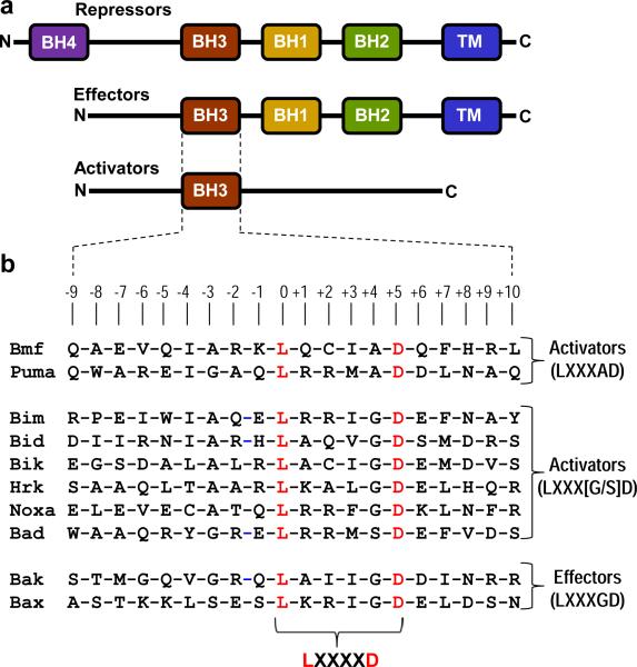
B-cell lymphoma protein 2 (Bcl2) apoptotic repressor carries out its function by virtue of its ability to bind to BH3 domains of various pro-apoptotic regulators in a highly promiscuous manner. Herein, we investigate the biophysical basis of such promiscuity of Bcl2 toward its cognate BH3 ligands. Our data show that although the BH3 ligands harboring the LXXXAD motif bind to Bcl2 with submicromolar affinity, those with the LXXX[G/S]D motif afford weak interactions. This implies that the replacement of alanine at the fourth position (A + 4)-relative to the N-terminal leucine (L0) within the LXXXAD motif-to glycine/serine results in the loss of free energy of binding. Consistent with this notion, the A + 4 residue within the BH3 ligands harboring the LXXXAD motif engages in key intermolecular van der Waals contacts with A149 lining the ligand binding groove within Bcl2, whereas A + 4G/S substitution results in the disruption of such favorable binding interactions. Of particular interest is the observation that although increasing ionic strength has little or negligible effect on the binding of high-affinity BH3 ligands harboring the LXXXAD motif, the binding of those with the LXXX[G/S]D motif in general experiences a varying degree of enhancement. This salient observation is indicative of the fact that hydrophobic forces not only play a dominant but also a universal role in driving the Bcl2-BH3 interactions. Taken together, our study sheds light on the molecular basis of the factors governing the promiscuous binding of Bcl2 to pro-apoptotic regulators and thus bears important consequences on the development of rational therapeutic approaches.

Keywords: binding thermodynamics; molecular dynamics; salt dependence; structural models.


Figures

Figure 1
Figure 1
An overview of Bcl2 family of proteins. (a) Structural organization of pro-survival (repressors) and pro-apoptotic (effectors and activators) regulators. The activators belong to the BH3-only proteins, where BH3 is the Bcl2 homology 3 domain. Examples of activators include Bad, Bid, Bik, Bim, Bmf, Hrk, Noxa and Puma. The effectors contain the BH3-BH1-BH2-TM modular architecture, where TM is the transmembrane domain located C-terminal to Bcl2 homology domains BH3, BH1 and BH2. Examples of effectors are Bak and Bax. The repressors are usually characterized by the BH4-BH3-BH1-BH2-TM modular organization, with an additional N-terminal Bcl2 homology 4 domain. Examples of repressors are Bcl2, BclXL, BclW, Mcl1 and Bfl1. (b) Amino acid sequence alignment of BH3 domains of various activators and effectors encoded by the human genome and employed in this study as ligands for Bcl2. Note that the absolutely conserved consensus leucine and aspartate residues within the LXXXXD motif shared by all BH3 domains are colored red. The numerals indicate the nomenclature used in this study to distinguish residues within and flanking the core LXXXXD motif relative to the consensus leucine, which is arbitrarily assigned zero.
Figure 2
Figure 2
Representative ITC isotherms for the binding of BH3 peptides of Puma (a), Bax (b) and Bad (c) to Bcl2 in Sodium phosphate buffer containing 100mM NaCl at 25°C and pH 7. Note that these BH3 peptides harbor LXXXAD (Puma), LXXXGD (Bax) and LXXXSD (Bad) motifs. The upper panels show raw ITC data expressed as change in thermal power with respect to time over the period of titration. In the lower panels, change in molar heat is expressed as a function of molar ratio of each BH3 peptide to Bcl2. The solid lines in the lower panels show non-linear least squares fit of data to a one-site binding model using ORIGIN as described previously (Wiseman et al., 1989; Bhat et al., 2012).
Figure 3
Figure 3
Inter-dependence of enthalpic (ΔH) and entropic (TΔS) contributions to the free energy (ΔG) for the binding of various BH3 peptides to Bcl2 in Sodium phosphate buffer containing 100mM NaCl at 25ΔC and pH 7. (a) TΔS-ΔH plot. (b) ΔH-ΔG plot. (c) TΔS-ΔG plot. Note that the Class I (red) and Class II (green) BH3 ligands display distinct thermodynamic behaviors that can be grouped together as indicated by the linear fit of appropriate data points (solid lines). Error bars were calculated from at least three independent measurements to one standard deviation.
Figure 4
Figure 4
Structural models of Bcl2 bound to BH3 peptides of Puma (a), Bax (b) and Bad (c). Note that these BH3 peptides harbor LXXXAD (Puma), LXXXGD (Bax) and LXXXSD (Bad) motifs. In each model, Bcl2 is shown in green and the corresponding BH3 peptide is colored yellow. The expanded views show sidechain moities of residues within Bcl2 and the corresponding BH3 peptide engaged in key intermolecular contacts in red and blue, respectively.
Figure 5
Figure 5
BH3 peptides undergo conformational changes upon binding to Bcl2 in Sodium phosphate buffer containing 100mM NaCl at pH 7. (a) Dependence of enthalpy (ΔH) on temperature (T) for the binding of Bcl2 to BH3 peptides harboring LXXXAD (Puma), LXXXGD (Bax) and LXXXSD (Bad) motifs. The solid lines through the data points represent linear fits. Error bars were calculated from at least three independent measurements to one standard deviation. (b) Dependence of free energy (ΔG) and the underlying enthalpic (ΔH) and entropic (TΔS) components on the total change in SASA (ΔSASAtotal) upon the binding of various BH3 peptides to Bcl2. Note that the Class I (red) and Class II (green) BH3 ligands display distinct thermodynamic behaviors that can be grouped together as indicated by the connecting of appropriate data points with solid lines. Error bars were calculated from at least three independent measurements to one standard deviation.
Figure 6
Figure 6
Effect of NaCl concentration on the binding, as measured by the binding constant (Kd), of Bcl2 to various BH3 peptides harboring LXXXAD and LXXX[G/S]D motifs in Sodium phosphate buffer at 25°C and pH 7. Note that the solid lines are used to connect various data points for clarity. Error bars were calculated from at least three independent measurements to one standard deviation.
Figure 7
Figure 7
Root mean square deviation (RMSD) and fluctuation (RMSF) of backbone atoms (N, Cα and C) obtained from MD analysis on the structural models of Bcl2 bound to various BH3 peptides harboring LXXXAD (Puma), LXXXGD (Bax) and LXXXSD (Bad) motifs. (a) RMSD of backbone atoms within each simulated structure relative to the initial modeled structure of Bcl2 bound to BH3 peptides of Puma (top panel), Bax (middle panel) and Bad (bottom panel) for the corresponding Bcl2-peptide complex (black), Bcl2 alone (green) and BH3 peptide alone (yellow) as a function of simulation time. (b) RMSF of backbone atoms averaged over the entire course of corresponding MD trajectory of Bcl2 bound to BH3 peptides of Puma (top panel), Bax (middle panel) and Bad (bottom panel) as a function of residue number within Bcl2. The shaded vertical rectangular box denotes residues located within the α1-α2 loop. (c) RMSF of backbone atoms averaged over the entire course of corresponding MD trajectory of Bcl2 bound to BH3 peptides of Puma (top panel), Bax (middle panel) and Bad (bottom panel) as a function of residue number within each peptide. The shaded vertical rectangular box denotes residues located within the LXXXXD motif.
Figure 8
Figure 8
Inter-atomic distances obtained from MD analysis on the structural models of Bcl2 bound to various BH3 peptides harboring LXXXAD (Puma), LXXXGD (Bax) and LXXXSD (Bad) motifs. (a) Distance between Cδ benzyl ring atoms of F153 (F153.Cδ1/δ2) within Bcl2 and Cδ methyl atoms of L0 (L0.Cδ1/δ2) within the BH3 peptides of Puma (top panel), Bax (middle panel) and Bad (bottom panel) as a function of simulation time (black). (b) Distance between Cζ atom of R146 (R146.Cζ) within Bcl2 and Cγ atom of D+5 (D+5.Cγ) within the BH3 peptides of Puma (top panel), Bax (middle panel) and Bad (bottom panel) as a function of simulation time (black). (c) Distances between Cβ atom of A149 (A149.Cβ) within Bcl2 and Cα atom of X+4 (X+4.Cα) within the BH3 peptides of Puma (top panel), Bax (middle panel) and Bad (bottom panel) as a function of simulation time (black), or between Cβ atom of A149 (A149.Cβ) within Bcl2 and Cβ atom of X+4 (X+4.Cβ) within the BH3 peptides of Puma (top panel) and Bad (bottom panel) as a function of simulation time (green). Note that X+4 is respectively alanine, glycine and serine within the BH3 peptides of Puma, Bax and Bad.

References

    1. Adams JM, Cory S. The Bcl-2 protein family: arbiters of cell survival. Science. 1998;281:1322–1326. - PubMed
    1. Berendsen HJC, Grigera JR, Straatsma TP. The Missing Term in Effective Pair Potentials. J Phys Chem. 1987;91:6269–6271.
    1. Bhat V, McDonald CB, Mikles DC, Deegan BJ, Seldeen KL, Bates ML, Farooq A. Ligand Binding and Membrane Insertion Compete with Oligomerization of the BclXL Apoptotic Repressor. J Mol Biol. 2012;416:57–77. - PMC - PubMed
    1. Boersma MD, Sadowsky JD, Tomita YA, Gellman SH. Hydrophile scanning as a complement to alanine scanning for exploring and manipulating protein-protein recognition: application to the Bim BH3 domain. Protein Sci. 2008;17:1232–1240. - PMC - PubMed
    1. Carson M. Ribbons 2.0. J Appl Crystallogr. 1991;24:958–961.

Publication types

MeSH terms