. 2014 Feb;13(1):151-77.
doi: 10.1007/s12311-013-0511-x.

Consensus paper: the cerebellum's role in movement and cognition

Affiliations

Consensus paper: the cerebellum's role in movement and cognition

Leonard F Koziol et al. Cerebellum. 2014 Feb.

Abstract

While the cerebellum's role in motor function is well recognized, the nature of its concurrent role in cognitive function remains considerably less clear. The current consensus paper gathers diverse views on a variety of important roles played by the cerebellum across a range of cognitive and emotional functions. This paper considers the cerebellum in relation to neurocognitive development, language function, working memory, executive function, and the development of cerebellar internal control models and reflects upon some of the ways in which better understanding the cerebellum's status as a "supervised learning machine" can enrich our ability to understand human function and adaptation. As all contributors agree that the cerebellum plays a role in cognition, there is also an agreement that this conclusion remains highly inferential. Many conclusions about the role of the cerebellum in cognition originate from applying known information about cerebellar contributions to the coordination and quality of movement. These inferences are based on the uniformity of the cerebellum's compositional infrastructure and its apparent modular organization. There is considerable support for this view, based upon observations of patients with pathology within the cerebellum.


Figures

Fig. 1
Fig. 1
a Diagram of the cerebro-cerebellar circuit. Feedforward limb: the corticopontine pathway (1) carries associative, paralimbic, sensory, and motor information from the cerebral cortex to the neurons in the ventral pons. The axons of these pontine neurons reach the cerebellar cortex via the pontocerebellar pathway (2). Feedback limb: the cerebellar cortex is connected with the deep cerebellar nuclei (3), which course through the midbrain in the vicinity of the red nucleus to terminate in the thalamus (the cerebello-thalamic projection, 4). The thalamic projection back to cerebral cortex (5) completes the feedback circuit. b Plane of section through the pons from which the rostrocaudal levels II through VIII are taken in the schematic (c). c Composite color-coded summary diagram illustrating the distribution within selected regions of the basis pontis of projections from association and paralimbic areas shown on medial, lateral, and orbital views of the cerebral hemisphere in the prefrontal (purple), posterior parietal (blue), superior temporal (red), and parastriate and parahippocampal regions (orange) and from motor, premotor, and supplementary motor areas (green). Other cerebral areas known to project to the pons are depicted in white. Cortical areas with no pontine projections are shown in yellow (from anterograde and retrograde studies) or gray (from retrograde studies). Dashed lines in the hemisphere diagrams represent sulcal cortices. Dashed lines in the pons diagrams represent pontine nuclei; solid lines depict corticofugal fibers. d Lateral view of monkey brain (top) shows the locations of viral tracer injections in the M1 arm, PMv arm, and prefrontal cortex areas 46 and 9. The resulting retrogradely labeled neurons in the cerebellar dentate nucleus (bottom) are indicated by solid dots. e Flattened representation of cerebellum to show the folia linked with M1 motor cortex (left) and prefrontal cortex area 46 (right) using viral tracers that travel in the anterograde direction (H129 strain of HSV1) and retrograde direction (rabies virus). ac reproduced and adapted from Schmahmann [62] and Schmahmann and Pandya [34, 35, 51]; d from Middleton and Strick [208]; e from Kelly and Strick [30]
Fig. 2
Fig. 2
Coronal slices through the cerebellum of a single individual showing topographic arrangement of fMRI activation patterns for tasks of finger tapping, color coded in red-orange; verb generation, blue; n-back, purple; mental rotation, green; and International Affective Picture Rating Scale, yellow. Left cerebellum is on the left, and coronal levels at y=−44, −56, −68, and −76 are shown. Activations are present in cerebellar lobules V, VI, Crus I, Crus II, VIIB, and VIII, as labeled. (Reproduced from Stoodley et al. [262]. Nomenclature of cerebellar lobules as in Schmahmann et al. [263])
Fig. 3
Fig. 3
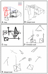
Visual spatial disintegration in the cerebellar cognitive affective syndrome. a Copy of the Rey figure by a 15-year-old boy with near-total cerebellar agenesis showing piecemeal performance rather than overall conceptual understanding of the figure. Diagram drawn using pencil, then black, blue, and red pen sequentially. b Delayed recall of the figure showing impaired recall and design. Performance on the Rey figure by a 6-year-old boy after resection of a left cerebellar cystic astrocytoma is shown in c copy, d immediate recall, and e delayed recall. Concept, design, and recall are impaired, with fragmentation of the image in (e) reminiscent of loosening of associations as may be seen in a psychiatric context. (a, b from Chheda et al. [63]. c, d from Levisohn et al. [61])
Fig. 4
Fig. 4
Schizophrenia patients (a) have significantly less cerebellar blood flow than healthy normals (b) during several cognitive tasks: (1) theory of mind, (2) recalling words, (3) recalling stories, and (4) emotion attribution. Representative images from each task were chosen to illustrate cerebellar recruitment during each of these cognitive tasks. Images follow radiological convention. Regions in red/yellow tones indicate positive peaks and regions with blue/purple indicate negative peaks. Each cognitive task is organized by two columns containing a threshold on the right (significant differences at the 0.005 level) and global blood flow column subdivided by slice orientation axial (top), sagittal (middle), and coronal (bottom) on the left. Statistical results are portrayed using the value of the associated t statistic, which is shown on the color bar on the right
Fig. 5
Fig. 5
Schematic illustrates the relationship between superior cerebellar lobule VI activity and verbal working memory performance. Neuroim-aging results from young, healthy adults performing a high vs. low load VWM task (149) showed that (a) activity in superior cerebellar lobule VI increased specifically in response to high load working memory demands, and (b) that this activity was inversely correlated to overall performance accuracy (r=−0.79). Increased lobule VI activity may reflect the temporal sequencing of motor traces representing inner speech. This strategy continues even when performance begins to decline, indicating an ongoing struggle to keep up with working memory demands. In (a) and (b), p<0.001–0.00001
Fig. 6
Fig. 6
Flattened view of cerebellar surface illustrating that the anterior lobe and intermediate parts of the posterior lobe are related to “motor and somatosensory functions,” whereas the lateral posterior cerebellum is related to “cognitive functions.” To orient properly to the anterior/posterior axis of the flattened view, the viewer should keep in mind that anterior/posterior refer to what is actually a substantially convex cerebellar surface (see smaller drawing to left). Arrows in (a) indicate difference between “motor” (note modularity of somatotopic maps at top and bottom) and “cognition” found in previous neuroimaging studies. Arrows at (b) indicate modularity within the lateral posterior cerebellum for two different cognitive functions. (Adapted from Imamizu et al. [179], p. 5461). Copyright 2003 by Hiroshi Imamizu. Reprinted with Permission
Fig. 7
Fig. 7
An example of embodied problem solving. During climbing competitions, before climbing an unknown route, athletes can pre-view it for some minutes; they typically mimic, imagine, and plan their future climbing movements (in the picture, they do this overtly, but this is not always the case). This is a complex problem solving, as (route settlers ensure that) the sequence of movements to reach the top of the wall is novel and far from trivial. It depends on goal state (the climbing hold to reach) and previous movements. Furthermore, it incorporates the athlete's embodied knowledge, as length of limbs, strength of fingers, affordances offered by the various kinds of climbing holds, possible or impossible kinematics, all constraint the problem solving process. Part of the athletes' skill is in the ability to anticipate relevant information (proprioceptive, body posture at critical points, how much force to use, etc.) prior to climbing, to form motor plans, and to maintain and refine them in memory before climbing. We reported in Pezzulo et al. [218] an advantage of expect climbers in a memory task (i.e., remembering sequences of holds in a route) but only when “climb-ability” constraints were respected (not when the sequences formed a nonclimbable route)
Fig. 8
Fig. 8
Schematic illustration of a cerebellar internal model learning spatiotemporal information. Top In eyeblink conditioning, the cerebellar internal model (drawing of the cerebellum in the middle) receives a spike train that is temporally constant (conditioned stimulus, left) and exerts another spike train that modulates in time maximally at the onset of an unconditioned stimulus (right). Bottom In phase adaptation of the vestibulo-ocular reflex, the internal model receives a spike train that modulates sinusoidally from semicircular canals and exerts another sinusoidally modulating spike train with a certain phase difference. In this way, an internal model is a general-purpose supervised learning machine of pairs of spatiotemporal signals. Horizontal bar, time axis; vertical bars, spikes

References

    1. Schmahmann JD. The cerebellum and cognition. Academic; London: 1997.
    1. Diamond A. Close interrelation of motor development and cognitive development and of the cerebellum and prefrontal cortex. Child Dev. 2000;71(1):44–56. - PubMed
    1. Baillieux H, Smet HJ, Paquier PF, De Deyn PP, Marien P. Cerebellar neurocognition: Insights into the bottom of the brain. Clin Neurol Neurosurg. 2008;110(8):763–73. - PubMed
    1. Kuper M, Dimitrova A, Thurling M, Maderwald S, Roths J, Elles HG, et al. Evidence for a motor and a non-motor domain in the human dentate nucleus—An fMRI study. Neuroimage. 2011;54(4):2612–22. - PubMed
    1. Molinari M, Chiricozzi FR, Clausi S, Tedesco AM, De LM, Leggio MG. Cerebellum and detection of sequences, from perception to cognition. Cerebellum. 2008;7(4):611–5. - PubMed

Publication types