

Review
doi: 10.1152/physiol.00007.2013.
Regulation of luminal acidification by the V-ATPase
Affiliations
- PMID: 23997191
- PMCID: PMC3768094
- DOI: 10.1152/physiol.00007.2013
Item in Clipboard
Review
Regulation of luminal acidification by the V-ATPase
Physiology (Bethesda).
2013 Sep.
Abstract
Specialized cells in the body express high levels of V-ATPase in their plasma membrane and respond to hormonal and nonhormonal cues to regulate extracellular acidification. Mutations in or loss of some V-ATPase subunits cause several disorders, including renal distal tubular acidosis and male infertility. This review focuses on the regulation of V-ATPase-dependent luminal acidification in renal intercalated cells and epididymal clear cells, which are key players in these physiological processes.
Conflict of interest statement
No conflicts of interest, financial or otherwise, are declared by the author(s).
Figures
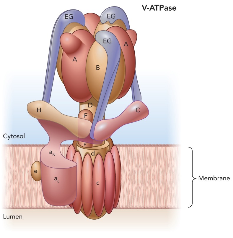
V-ATPase subunit composition and organization Subunits A, B, C, D, E, F, G, and H form the V1 cytosolic domain, and subunits a, c, d, and e form the V0 transmembrane domain. ATP catalytic sites are located at the B/A subunit interfaces. Protons are transported between subunit a and the proteolipid c-ring. The V-ATPase rotor is formed by subunits D, F, and d and the c-ring. Three EG complexes together with subunits C and H form a stator, which is connected to the AB hexamer and the large transmembrane subunit a. The function of subunit e is still unknown. Image is based on data from Refs. and 88.
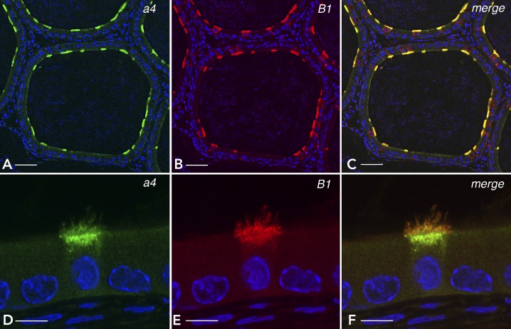
Localization of the V-ATPase B1 and a4 subunit isoforms in epididymal clear cells The distal region (cauda) of rat epididymis was immunofluorescently labeled with antibodies against a4 (A and D: green) and B1 (B and E: red). C and F: merged images from A and B, and D and E, respectively. B1 and a4 colocalize in the apical membrane and subapical vesicles in clear cells exclusively. Nuclei and sperm are labeled in blue with DAPI. Bars = 50 μm (A, B, C) and 5 μm (D, E, F).
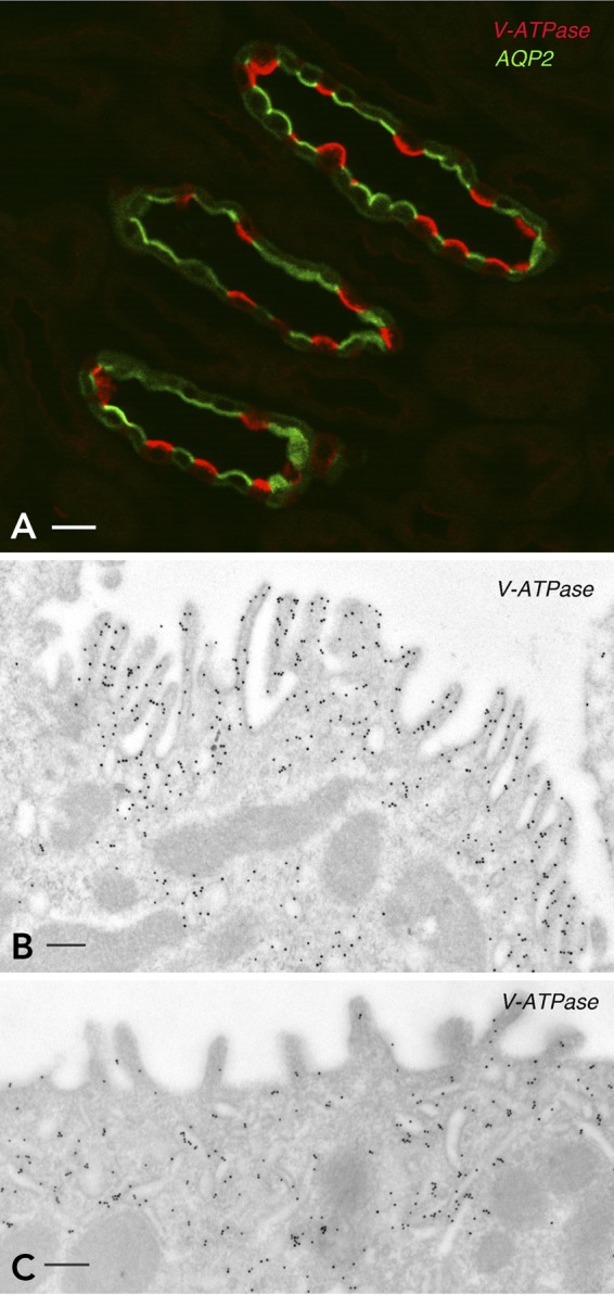
Expression of the V-ATPase in kidney intercalated cells A: the renal inner stripe of the outer medulla was immunofluorescently labeled for the V-ATPase B1 subunit (red) and AQP2 (green). The V-ATPase and AQP2 are located apically in intercalated cells and principal cells, respectively. B and C: immunogold electron microscopy labeling of the apical pole of two intercalated cells for the V-ATPase A subunit. B: an activated IC with numerous apical microvilli that contain a high density of V-ATPase-associated gold particles. C: a resting IC with fewer and less developed microvilli. Most V-ATPase-gold particles are located in subapical tubulo-vesicles.

Immunofluorescence labeling of CCs from the cauda epididymis Immunofluorescence labeling of CCs from the cauda epididymis after in vivo luminal perfusion with a control solution adjusted to the resting pH of 6.6 (A) or with an activating solution containing bicarbonate and the cAMP permeant analog cpt-cAMP (B). Horseradish peroxidase (HRP) was added to both solutions to label endosomes (red). Epididymis sections were labeled for the V-ATPase B1 subunit (green). Arrows indicate the frontier between the apical cytoplasm and the base of microvilli. Short microvilli labeled for the V-ATPase are detected in the resting CC shown in A. The yellow color in A shows colocalization of V-ATPase in HRP-containing endosomes located in the subapical pole of clear cells. In the activated CC shown in B, the V-ATPase is mainly located in longer and more numerous microvilli (green), and it is absent from the apical endosomes that contain the internalized HRP (red). Scale bars = 5 μm. Figure reproduced from Ref. with permission from the American Society of Andrology.
References
-
- Al-Awqati Q. Plasticity in epithelial polarity of renal intercalated cells: targeting of the H+-ATPase and band 3. Am J Physiol Cell Physiol 270: C1571–C1580, 1996 - PubMed
-
- Alzamora R, Thali RF, Gong F, Smolak C, Li H, Baty CJ, Bertrand CA, Auchli Y, Brunisholz RA, Neumann D, Hallows KR, Pastor-Soler NM. PKA regulates vacuolar H+-ATPase localization and activity via direct phosphorylation of the a subunit in kidney cells. J Biol Chem 285: 24676–24685, 2010 - PMC - PubMed
-
- Banerjee A, Shih T, Alexander EA, Schwartz JH. SNARE proteins regulate H+-ATPase redistribution to the apical membrane in rat renal inner medullary collecting duct cells. J Biol Chem 274: 26518–26522, 1999 - PubMed
-
- Beaulieu V, Da Silva N, Pastor-Soler N, Brown CR, Smith PJ, Brown D, Breton S. Modulation of the actin cytoskeleton via gelsolin regulates vacuolar H+ATPase (V-ATPase) recycling. J Biol Chem 280: 8452–8463, 2005 - PubMed
Publication types
MeSH terms
Substances
Grants and funding
- DK-042956/DK/NIDDK NIH HHS/United States
- DK-097124/DK/NIDDK NIH HHS/United States
- P30 DK043351/DK/NIDDK NIH HHS/United States
- DK43351/DK/NIDDK NIH HHS/United States
- DK-038452/DK/NIDDK NIH HHS/United States
- R56 DK042956/DK/NIDDK NIH HHS/United States
- R37 DK042956/DK/NIDDK NIH HHS/United States
- R01 DK042956/DK/NIDDK NIH HHS/United States
- R01 DK097124/DK/NIDDK NIH HHS/United States
- P30 DK057521/DK/NIDDK NIH HHS/United States
- R01 HD040793/HD/NICHD NIH HHS/United States
- HD-040793/HD/NICHD NIH HHS/United States
- P01 DK038452/DK/NIDDK NIH HHS/United States
- DK57521/DK/NIDDK NIH HHS/United States
LinkOut - more resources
Full Text Sources
Other Literature Sources