. 2013 Sep;257(17-18):2565-2588.
doi: 10.1016/j.ccr.2013.02.007.

Designing functional metalloproteins: from structural to catalytic metal sites

Affiliations

Designing functional metalloproteins: from structural to catalytic metal sites

Melissa L Zastrow et al. Coord Chem Rev. 2013 Sep.

Abstract

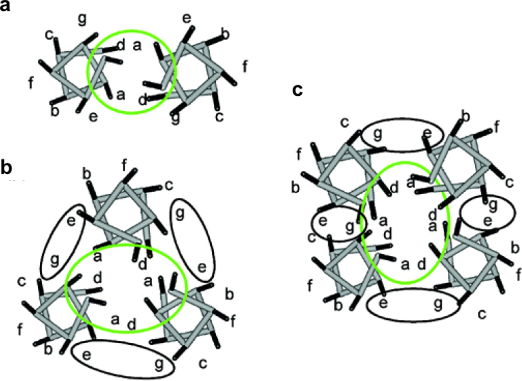
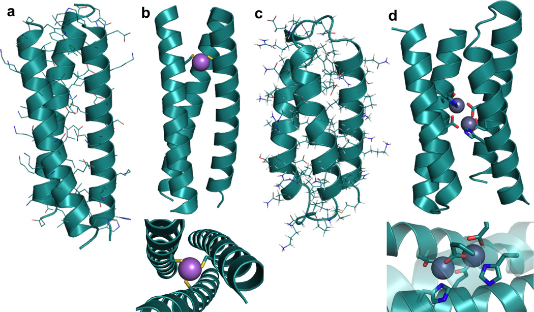
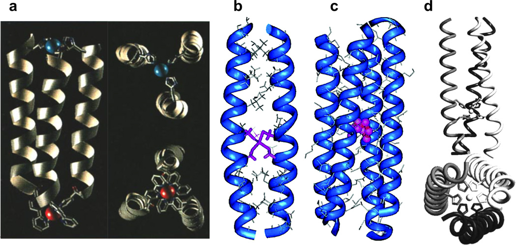
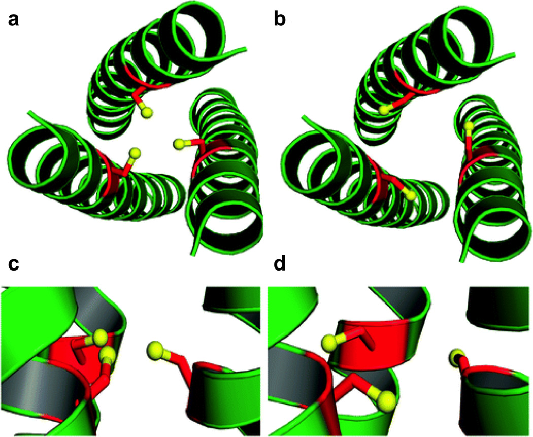
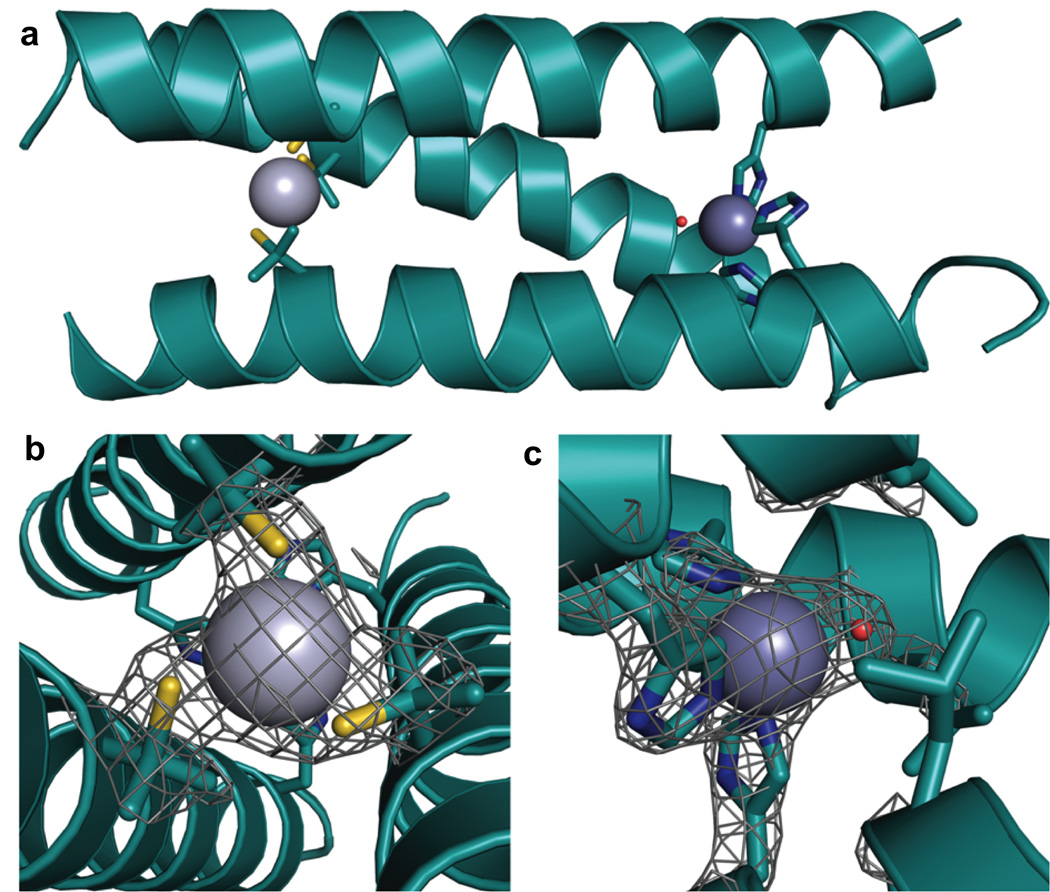
Metalloenzymes efficiently catalyze some of the most important and difficult reactions in nature. For many years, coordination chemists have effectively used small molecule models to understand these systems. More recently, protein design has been shown to be an effective approach for mimicking metal coordination environments. Since the first designed proteins were reported, much success has been seen for incorporating metal sites into proteins and attaining the desired coordination environment but until recently, this has been with a lack of significant catalytic activity. Now there are examples of designed metalloproteins that, although not yet reaching the activity of native enzymes, are considerably closer. In this review, we highlight work leading up to the design of a small metalloprotein containing two metal sites, one for structural stability (HgS3) and the other a separate catalytic zinc site to mimic carbonic anhydrase activity (ZnN3O). The first section will describe previous studies that allowed for a high affinity thiolate site that binds heavy metals in a way that stabilizes three-stranded coiled coils. The second section will examine ways of preparing histidine rich environments that lead to metal based hydrolytic catalysts. We will also discuss other recent examples of the design of structural metal sites and functional metalloenzymes. Our work demonstrates that attaining the proper first coordination geometry of a metal site can lead to a significant fraction of catalytic activity, apparently independent of the type of secondary structure of the surrounding protein environment. We are now in a position to begin to meet the challenge of building a metalloenzyme systematically from the bottom-up by engineering and analyzing interactions directly around the metal site and beyond.

Keywords: Hg thiolates; Zn hydrolase; de novo metalloprotein design.


Figures

Figure 1
Figure 1
Helical wheel diagrams for parallel (a) two-, (b) three-, and (c) four-stranded coiled coils. Figure was reproduced with permission from ref. [53]. Copyright 2004 American Chemical Society.
Figure 2
Figure 2
Structures of representative de novo designed α-helical coiled coils and bundles. (a) X-ray structure of a three-stranded coiled coil, CoilSer generated from PDB 1COS [50], (b) X-ray structure of a metal-bound three-stranded coiled coil, [AsIII]S(CSL9C)3 generated from PDB 2JGO [68] with side-on view (top) and top-down view (bottom); As-S metal-ligand bond distances all 2.3 Å, (c) NMR structure of a three-helix α-helical bundle protein generated from PDB 2A3D [73], (d) X-ray structure of a dinuclear metal site in a designed four-helix bundle protein (diZnII-DF1) generated from PDB 1EC5 [11] with side-on full view (top) and close-up view of metal site (bottom); All metal-ligand bond distances are in the range 1.8–2.1 Å.
Figure 3
Figure 3
Examples of metal-stabilized α-helical structures described in this section. (a) Computer-generated model of the parallel three-helix bundle CuIIRuII metalloprotein with full side-on view (left) and close-up views of each metal site (right). Figure was reproduced with permission from ref. [113]. Copyright WileyVCH Verlag GmbH & Co. KGaA. (b) Energy-minimized computer model of the CdII-bridged C16C19 peptide dimer (2SCC) with CdII bound in a tetrahedral geometry. Figure was reproduced with permission from ref. [7]. Copyright 2006 American Chemical Society. (c) Computer-generated model of the tetrameric CuI-C16C19-GGY metalloprotein (4SCC). Figure was reproduced with permission from ref. [7]. Copyright 2006 American Chemical Society. (d) Model of the NiII-His6 complex of IZ-3adH (3SCC) with full side-on view (top) and a bottom-up view from the C-termini (bottom). Figure was reproduced with permission from ref. [109]. Copyright 1998 American Chemical Society.
Figure 4
Figure 4
Ribbon diagrams of the X-ray crystal structure of apo(CSL9C)3 showing the orientation of the Cys ligands. The Cys side chains are shown as red sticks with thiol groups colored yellow. Top-down views from the N-termini show (a) the orientation of the major conformer with all Cys side chains pointing towards the interior of the trimer and (b) the minor conformer with Cys side chains pointing towards the helical interface. Side views further demonstrate the flexibility of this site. (c) In the major conformer, the thiol groups point towards the N-termini and (d) in the minor conformer, the thiol groups point towards the C-termini. Figure was reproduced with permission from ref. [67]. Copyright 2010 American Chemical Society.
Figure 5
Figure 5
Ribbon diagrams of the X-ray crystal structure of apo(CSL19C)3 showing the orientation of the Cys ligands. The Cys side chains are shown as red sticks with thiol groups colored yellow. The top-down view from the N-termini shows (a) that two of the Cys ligands point towards the interior of the coiled coil and the third toward the helical interface. The side view demonstrates that two thiol groups point towards the C-termini while the third is almost perpendicular to the helical axis. Figure was reproduced with permission from ref. [67]. Copyright 2010 American Chemical Society.
Figure 6
Figure 6
Ribbon diagrams of the X-ray crystal structures of apo(CSL16Pen)3 and apo(CSL16d-Pen)3 displaying the orientation of the Pen side chain. The l-(purple) and d-Pen (blue) side chains are shown in stick form with the thiol group colored orange. Side views show the (a) l-Pen residues oriented toward the N-termini and the (b) d-Pen residues oriented toward the C-termini. Top down stereo views from the N-termini are shown and display (c) l-Pen residues oriented toward the interior of the coiled coil and (b) d-Pen residues oriented toward the helical interface. Figure was reproduced with permission from ref. [122]. Copyright © 2009 WILEYVCH Verlag GmbH & Co. KGaA, Weinheim.
Figure 7
Figure 7
Pymol models of possible metal-binding geometries in a 3SCC. The metal-binding ligands are Cys residues and the metal is colored purple. (a) side-on view (top) and top-down view (bottom) of two-coordinate or linear geometry, (b) side-on view (top) and top-down view (bottom) of three-coordinate trigonal planar geometry, c) side-on view (top) and top-down view (bottom) of four-coordinate tetrahedral geometry, (d) side-on view of five-coordinate trigonal bipyramidal geometry, and (e) side-on view of six-coordinate octahedral metal geometry. Models are generated using the crystal structure of [AsIII]S(CSL9C)3 PDB 2JGO [68].
Figure 8
Figure 8
Species present at different TRIL9C/HgII ratios and pH values. Figure was reproduced from ref. [199] with permission of the copyright holders. Figure 9. Linear-free energy correlation between folding preferences of the peptides in the absence of metal to the binding of a third strand of peptide to a divalent HgIIS2 species. Figure was reproduced with permission from ref. [200]. Copyright © 2007 WILEYVCH Verlag GmbH & Co. KGaA, Weinheim.
Figure 9
Figure 9
Linear-free energy correlation between folding preferences of the peptides in the absence of metal to the binding of a third strand of peptide to a divalent HgIIS2 species. Figure was reproduced with permission from ref. [200]. Copyright 2005 American Chemical Society.
Figure 10
Figure 10
Stepwise aggregation-deprotonation (StepAD) mechanism for the encapsulation of HgII by the 3SCC, (BabyL9C)3. HgII reacts in a fast step to form [HgII]S(BabyL9C)2. Formation of [HgII]S(BabyL9C)2(H-BabyL9C) is the rate-limiting association and, depending on pH, rapidly converts to [HgII]S(BabyL9C)3. Figure was reproduced from ref. [125] with permission of the copyright holders.
Figure 11
Figure 11
Possible mechanisms for insertion of HgII into the folded peptides. Figure was reproduced with permission from ref. [53]. Copyright 2004 American Chemical Society.
Figure 12
Figure 12
Ribbon diagrams of the X-ray crystal structure of [HgII]S[ZnII(OH2/OH)]N(CSL9PenL23H)3n+ at pH 8.5. The Pen and His side chains are shown in stick form (sulfur = yellow; nitrogen = blue; oxygen = red). (a) One of two trimers found in the asymmetric unit of the crystal structure. (b) Top-down view of the structural trigonal thiolate site, HgIIS3, confirming the proposed structure of HgII in Cys-containing TRI. (c) Side view of the tetrahedral catalytic site, ZnIIN3O, which closely mimics carbonic anhydrase and matrix metalloproteinase active sites. Figure was reproduced from ref. [70] with permission of the copyright holders.
Figure 13
Figure 13
Guanidine hydrochloride denaturation titrations represented by the molar ellipticity values [Θ] at 222 nm versus denaturant concentration for TRIL23H (■), TRIL9CL23H (●), and TRIL9CL23H + 1/3 HgII (▲). Figure was reproduced from ref. [70] with permission of the copyright holders.
Figure 14
Figure 14
Comparison of the size of the active site cavities of (a) modeled His3 site using the structure of [AsIII]S(CSL9C)3 (PDB 2JGO)[68] and (b) the actual structure containing the His3 site, [HgII]S[ZnII(OH2/OH)]N(CSL9PenL23H)3 n+ (PDB 3PBJ) [70]. (c) Overlay of the two sites with the model in gray and the actual structure in cyan.
Figure 15
Figure 15
Overlay of the ZnIIN3O site in [HgII]S[ZnII(OH2/OH)]N(CSL9PenL23H)3 n+ (cyan, PDB 3BPJ) with the active site of human CAII (tan, PDB 2CBA). The solvent molecule of [HgII]S[ZnII(OH2/OH)]N(CSL9PenL23H)3 n+ is shown in red and that for CA lies directly underneath. Figure was reproduced from ref. [70] with permission of the copyright holders.
Figure 16
Figure 16
X-ray crystal structure of MID1-zinc, a designed metal-mediated protein interface. The red mesh represents the active site cleft above the open coordination site of the ZnHis3 metal site. Figure was reproduced with permission from ref. [16]. Copyright 2012 American Chemical Society.
Figure 17
Figure 17
Model of [CuI/II]N(TRIL23H)3+/2+ based on the structure of [HgII]S[ZnII(OH2/OH)]N(CSL9PenL23H)3n+ (PDB 3PBJ). (a) side view of the model, (b) Top-down view from the N-termini of the metal sites with the ZnII(H2O)(His)3 site in light grey superimposed on the Type 2 CuII(OH2)(His)3 site in R. sphaeroides NiR (PDB 2DY2) in dark grey. (c) side view of the superimposed metal sites, as in b. Figure was reproduced from ref. [162] with permission of the copyright holders.
Figure 18
Figure 18
Helix-wheel representation of the antiparallel four-stranded coiled coil structure surrounding the active site of a diiron protein. Residue positions are labeled according to the heptad repeat generally applied to coiled coils. Figure was reproduced from ref. [11] with permission of the copyright holders.
Figure 19
Figure 19
Ribbon diagrams of the structures of metal-bound and apo-DF1. (a) The X-ray crystal structure of di-ZnII-DF1 (PDB 1EC5) [11]. (b) The X-ray crystal structure of di- MnII-DF1 (PDB 1OVR) [177]. (c) The NMR structure of apo-DF1 (PDB 1NVO) [178]. The helix-loop-helix motif in front of the ligands is shown as transparent to give an improved view.
Figure 20
Figure 20
2Fo-Fc electron density maps of the dinuclear metal-binding site of di-MnIIL13G- DF1 in the dimer (one of four in the asymmetric unit) with terminal waters bound to each metal (top) and in one of the dimers with a bridging water (bottom). Figure was reproduced from ref. [82] with permission of the copyright holders.
Figure 21
Figure 21
Comparison of DF1 and DF3 structures. (a) Crystal structure of di-ZnII-DF1 (PDB 1EC5). (b) Surface representation of the crystal structure of di-ZnII-DF1 to display the accessibility to the dimetal site. (c) NMR structure of di-ZnII-DF3 (PDB 2KIK). (d) Surface representation of the NMR structure of di-ZnII-DF3 to show the increased accessibility to the active site over DF1. The different residues in position 9 (lime) and 13 (cyan) are highlighted. Metal ions are colored magenta. Figure was reprinted by permission from Macmillan Publishers Ltd: Nature Chemical Biology [81], copyright 2009.
Figure 22
Figure 22
Ribbon diagram showing the structure of the 3His-G2DFsc variant (PDB 2LFD) with the added metal-binding His residue (H100) and supporting mutations (I37N, L81H). Although kinetic data is reported for the 3His-G4DFsc variant, the structure shown here contains only two Gly mutations because of increased stability during data collection (this variant also showed N-oxygenase activity, albeit slower than the G4-variant). Figure was reprinted by permission from Macmillan Publishers Ltd: Nature Chemistry [80], copyright 2012.
Figure 23
Figure 23
Proposed reaction scheme for N-hydroxylation of p-anisidine. Figure was reprinted by permission from Macmillan Publishers Ltd: Nature Chemistry [80], copyright 2012.
Figure 24
Figure 24
Representation of structure-selective peptide modification by a ruthenium-bound peptide catalyst. 1 is the diazo reagent. X is the amino acid to be modified (Trp is discussed in the text). Figure was reproduced from ref. [195] with permission of The Royal Society of Chemistry.

References

    1. Holm RH, Kennepohl P, Solomon EI. Chem. Rev. 1996;96:2239. - PubMed
    1. Ragsdale SW. Chem. Rev. 2006;106:3317. - PubMed
    1. Dieckmann GR, McRorie DK, Lear JD, Sharp KA, DeGrado WF, Pecoraro VL. J. Mol. Biol. 1998;280:897. - PubMed
    1. Kaplan J, DeGrado WF. Proc. Natl. Acad. Sci. U.S.A. 2004;101:11566. - PMC - PubMed
    1. DeGrado WF, Raleigh DP, Handel T. Curr. Opin. Struct. Biol. 1991;1:984.

LinkOut - more resources