. 2012 Sep;29(3):155-60.
doi: 10.1055/s-0032-1326923.

Bronchial artery embolization

Affiliations

Bronchial artery embolization

Jonathan Lorenz et al. Semin Intervent Radiol. 2012 Sep.

Abstract

Hemoptysis represents a significant clinical entity with high morbidity and potential mortality. Most hemorrhages from a bronchial source arise in the setting of chronic inflammatory diseases. Medical management (in terms of resuscitation and bronchoscopic interventions) and surgery have severe limitations in these patient populations. Embolization procedures represent the first-line treatment for hemoptysis arising from a bronchial arterial source. This article discusses anatomical and technical considerations, as well as outcomes and complications, in the setting of bronchial arterial embolization in the treatment of hemoptysis.

Keywords: bronchial artery; embolization; hemoptysis; interventional radiology.


Figures

Figure 1
Figure 1
Frontal chest radiograph reveals extensive, bilateral bronchiectasis, particularly prominent in the right apex (arrow).
Figure 2
Figure 2
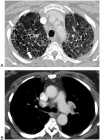
(A) Contrast-enhanced computed tomography (CT), lung windows, better depicts diffuse bilateral bronchiectasis, most prominent in the right upper lobe (arrows). (B) Contrast-enhanced CT, soft tissue windowing, demonstrates a right intercostobronchial trunk (arrow) that was targeted for catheterization.
Figure 3
Figure 3
(A) Aortogram, early arterial phase frontal projection, demonstrates a faint bronchial artery superimposed over the middescending thoracic aorta (open black arrow) as well as a prominent branch (white arrow) of the internal mammary artery superimposed over the right upper lobe. (B) Selective arteriography of the right intercostobronchial trunk shown in Figure 2A demonstrates supply to several intercostal arteries as well as a focus of hypervascularity (arrows) in the right lateral apex. This vessel was embolized with 500 to 700 micron particles injected through a microcatheter. (C) Selective arteriography of the right internal mammary artery demonstrates an enlarged branch (arrows) supplying the same hypervascular region previously embolized. (D) The internal mammary arterial branch was selectively catheterized with a microcatheter (arrow) and embolized with 500 to 700 micron particles. (E) Selective arteriography of the right internal mammary artery postembolization demonstrates stasis of this branch after embolization.
Figure 4
Figure 4
Computed tomography angiography with three-dimensional reconstruction of a patient with massive hemoptysis demonstrates a right bronchial artery (arrowhead) in its typical location, originating from the middescending aorta near the left mainstem bronchus (not shown). Three additional bronchial arteries (arrows) originate anomalously from the aortic arch, which is the most common source of anomalous bronchial supply.
Figure 5
Figure 5
(A) Frontal radiograph of a patient with massive hemoptysis demonstrates marked diffuse bilateral bronchiectasis. The location and most likely source of the bleeding is difficult to discern. (B) Contrast-enhanced computed tomography (CT), lung windowing and coronal reconstruction, demonstrates the most prominent foci of cystic bronchiectasis to reside in the right lower lobe. (C) Bronchial angiography, midarterial phase, demonstrates a hypervascular region corresponding to the bronchiectasis noted on the CT.
Figure 6
Figure 6
(A) Images from a patient with massive hemoptysis illustrates why large vessel occlusion is not appropriate in these cases. Selective brachiocephalic arteriography, early arterial phase, demonstrates hypertrophied branches of the thyrocervical trunk (arrow). (B) Subclavian artery injection performed near the origin of the thyrocervical trunk. Markedly enlarged collateral branch from the thyrocervical branch(es) (arrow) reconstitute a surgically clipped main bronchial artery (arrowhead). (C) The reconstituted bronchial artery noted in (B) supplies a region of hypervascularity (arrow) in the right lung.

References

    1. Rémy J, Arnaud A, Fardou H, Giraud R, Voisin C. Treatment of hemoptysis by embolization of bronchial arteries. Radiology. 1977;122(1):33–37. - PubMed
    1. Hartmann I J, Remy-Jardin M, Menchini L, Teisseire A, Khalil C, Remy J. Ectopic origin of bronchial arteries: assessment with multidetector helical CT angiography. Eur Radiol. 2007;17(8):1943–1953. - PubMed
    1. Bommart S, Bourdin A, Giroux M F. Transarterial ethylene vinyl alcohol copolymer visualization and penetration after embolization of life-threatening hemoptysis: technical and clinical outcomes. Cardiovasc Intervent Radiol. 2012;35(3):668–675. - PubMed
    1. Baltacioğlu F, Cimşit N C, Bostanci K, Yüksel M, Kodalli N. Transarterial microcatheter glue embolization of the bronchial artery for life-threatening hemoptysis: technical and clinical results. Eur J Radiol. 2010;73(2):380–384. - PubMed
    1. Cremaschi P, Nascimbene C, Vitulo P. et al.Therapeutic embolization of bronchial artery: a successful treatment in 209 cases of relapse hemoptysis. Angiology. 1993;44(4):295–299. - PubMed