. 2012 Sep;29(3):204-17.
doi: 10.1055/s-0032-1326931.

Inferior vena cava filtration in the management of venous thromboembolism: filtering the data

Affiliations

Inferior vena cava filtration in the management of venous thromboembolism: filtering the data

Christopher Molvar. Semin Intervent Radiol. 2012 Sep.

Abstract

Venous thromboembolism (VTE) is a common cause of morbidity and mortality. This is especially true for hospitalized patients. Pulmonary embolism (PE) is the leading preventable cause of in-hospital mortality. The preferred method of both treatment and prophylaxis for VTE is anticoagulation. However, in a subset of patients, anticoagulation therapy is contraindicated or ineffective, and these patients often receive an inferior vena cava (IVC) filter. The sole purpose of an IVC filter is prevention of clinically significant PE. IVC filter usage has increased every year, most recently due to the availability of retrievable devices and a relaxation of thresholds for placement. Much of this recent growth has occurred in the trauma patient population given the high potential for VTE and frequent contraindication to anticoagulation. Retrievable filters, which strive to offer the benefits of permanent filters without time-sensitive complications, come with a new set of challenges including methods for filter follow-up and retrieval.

Keywords: complications; deep vein thrombosis; inferior vena cava (IVC) filter; pulmonary embolism; trauma; venous thromboembolism.


Figures

Figure 1
Figure 1
A 45-year-old female trauma victim with contraindication to anticoagulation. (A) Preplacement vena cavogram with catheter placed via a right internal jugular vein approach. No intracaval thrombus or variant venous anatomy is present, and bilateral renal vein inflow is noted. (B) Technically successful infrarenal placement of a Celect inferior vena cava filter (Cook Medical Inc., Bloomington, IN). Case continues in Figs. 2 and 3.
Figure 2
Figure 2
Same patient as Fig. 1. Vena cavogram 2 months after inferior vena cava filter placement demonstrates trapped thrombus in the cone of the filter in two projections (arrows). (A) Anterior posterior and (B) oblique projection. Although trapped thrombus volume is ∼25% of the filter conical volume, the decision was made to leave the filter in place with continued anticoagulation and repeat vena cavogram in 1 month. Case continues in Fig. 3.
Figure 3
Figure 3
Same patient as Figs. 1 and 2. (A) Follow-up vena cavogram 3 months after filter placement shows resolution of filter-trapped thrombus without intracaval thrombus. The inferior vena cava (IVC) filter was removed using a loop snare. (B) Image of filter with retrieval hook engaged by a loop snare as a jugular sheath is advanced, collapsing the filter. (C) Spot image documented complete removal of the IVC filter.
Figure 4
Figure 4
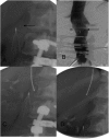
A 92-year-old woman with metastatic pancreatic cancer with recent pulmonary embolism despite therapeutic anticoagulation. (A) Vena cavogram via right internal jugular vein approach demonstrates a large filling defect in the caudal aspect of the inferior vena cava (IVC) consistent with thrombus. No variant venous anatomy was present. (B) Technically successful placement of infrarenal Vena Tech IVC filter, cephalad to the caval thrombus.
Figure 5
Figure 5
A 28-year-old man with low back pain and factor V Leiden deficiency with history of venous thromboembolism. Preoperative placement of a Recovery inferior vena cava (IVC) filter (Bard, Tempe, AZ) was performed. In the postoperative period with the patient ambulatory and therapeutic on anticoagulation, IVC filter removal was attempted. (A) Vena cavogram with pigtail catheter placed via a right internal jugular vein approach shows large nonocclusive thrombus trapped in the interstices of the filter. Anticoagulation was continued with attempted filter retrieval in 1 month. (B) A 1-month follow-up vena cavogram in an oblique projection demonstrates resolution of filter trapped thrombus and no intracaval thrombus. As such, the filter was removed. (C) Postretrieval vena cavogram demonstrates normal caliber IVC without complication.
Figure 6
Figure 6
A 52-year-old woman with prophylactic Recovery filter (Bard, Tempe, AZ), placed prior to spinal surgery; the patient developed chest pain several months after surgery. (A) Coronary angiogram with intracardiac inferior vena cava filter strut (arrow). (B) Left ventriculogram with additional filter strut in the right lung (arrow). The patient underwent operative removal of intracardiac filter strut with resolution of chest pain. Patient was then scheduled for IVC filter retrieval. Case continues in Figs. 7 and 8. Case courtesy of Marc Borge, MD.
Figure 7
Figure 7
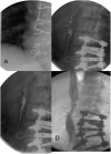
Same patient as Fig. 6. (A) Spot image prior to retrieval procedure shows filter with missing primary struts, along with an additional displaced strut (arrow). (B) Vena cavogram via a right common femoral approach demonstrates an intracaval location of a displaced strut (arrow). (C) Displaced strut is engaged with a loop snare and (D) pulled free from Recovery filter. Case continues in Fig. 8. Case courtesy of Marc Borge, MD.
Figure 9
Figure 9
Indwelling Günther Tulip inferior vena cava (IVC) filter (Cook Medical Inc., Bloomington, IN) is present in a patient therapeutic on anticoagulation. (A) Vena cavogram via a right internal jugular vein approach demonstrates an infrarenal filter with several struts projected minimally outside the contrast-opacified IVC. No intracaval thrombus or filter-trapped thrombus is noted. The filter was successfully removed. (B) Postremoval vena cavogram demonstrates contrast extravasation (arrow) at the level of previous filter-protruding struts. The patient was asymptomatic and vital signs were stable. (C) Resolution of contrast extravasation on 10-minute delayed vena cavogram.
Figure 8
Figure 8
Same patient as Figs. 6 and 7. (A) Spot image showing snared filter strut and intrapulmonary strut (arrow). (B, C) Recovery cone is seen engaging the filter and then collapsing it into a jugular sheath. (D) Postretrieval vena cavogram demonstrates irregular narrowing at site of previous filter consistent with spasm and/or scarring. Case courtesy of Marc Borge, MD.
Figure 10
Figure 10
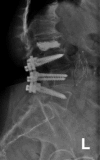
An 80-year-old woman status post spinal surgery and kyphoplasty. Fractured indwelling TrapEase filter (Cordis, Bridgewater, NJ) is seen on lumbar spine radiograph. The patient was asymptomatic.

References

    1. Anderson F A Jr, Zayaruzny M, Heit J A, Fidan D, Cohen A T. Estimated annual numbers of US acute-care hospital patients at risk for venous thromboembolism. Am J Hematol. 2007;82(9):777–782. - PubMed
    1. Tsai A W, Cushman M, Rosamond W D, Heckbert S R, Polak J F, Folsom A R. Cardiovascular risk factors and venous thromboembolism incidence: the longitudinal investigation of thromboembolism etiology. Arch Intern Med. 2002;162(10):1182–1189. - PubMed
    1. Kinney T B. Inferior vena cava filters. Semin Intervent Radiol. 2006;23(3):230–239. - PMC - PubMed
    1. Angel L F Tapson V Galgon R E Restrepo M I Kaufman J Systematic review of the use of retrievable inferior vena cava filters J Vasc Interv Radiol 201122111522–1530., e3 - PubMed
    1. Tapson V F. Acute pulmonary embolism. N Engl J Med. 2008;358(10):1037–1052. - PubMed