. 2013 Jun;8(3):290-312.
doi: 10.1016/j.nantod.2013.04.007.

Nonporous Silica Nanoparticles for Nanomedicine Application

Affiliations

Nonporous Silica Nanoparticles for Nanomedicine Application

Li Tang et al. Nano Today. 2013 Jun.

Abstract

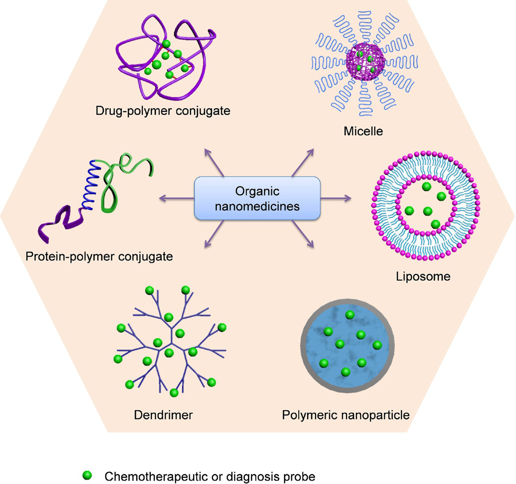
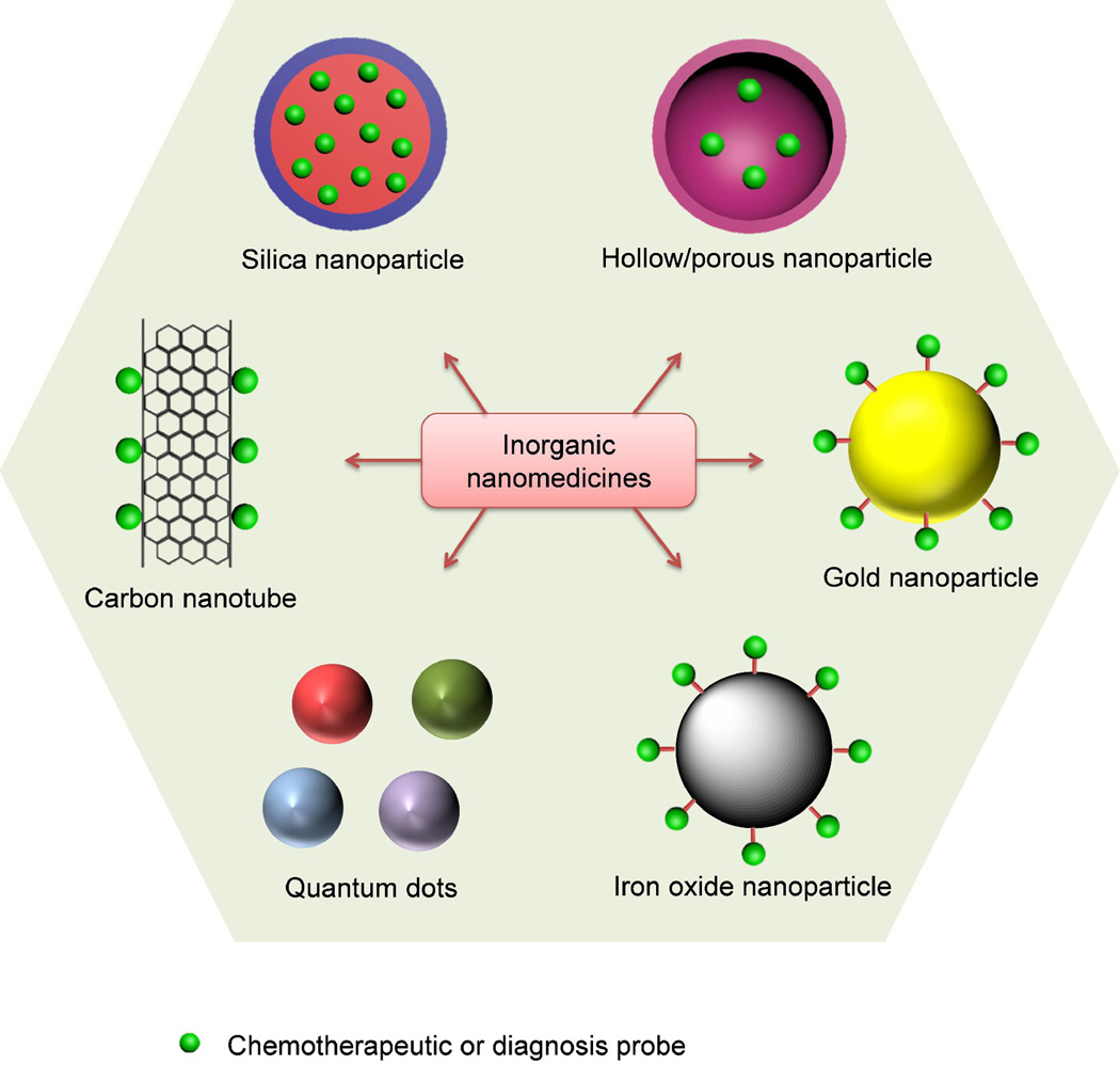
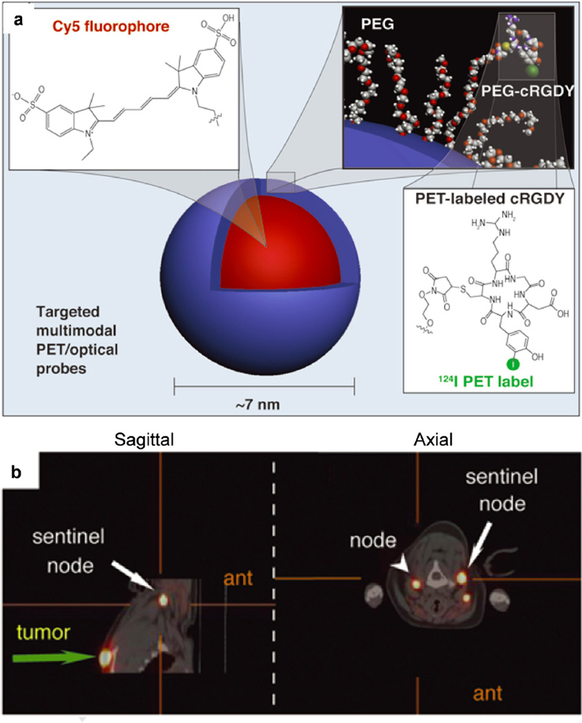
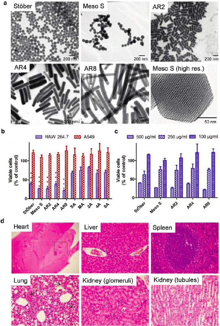
Nanomedicine, the use of nanotechnology for biomedical applications, has potential to change the landscape of the diagnosis and therapy of many diseases. In the past several decades, the advancement in nanotechnology and material science has resulted in a large number of organic and inorganic nanomedicine platforms. Silica nanoparticles (NPs), which exhibit many unique properties, offer a promising drug delivery platform to realize the potential of nanomedicine. Mesoporous silica NPs have been extensively reviewed previously. Here we review the current state of the development and application of nonporous silica NPs for drug delivery and molecular imaging.

Keywords: Nanomedicine; biomaterials; drug delivery; gene delivery; molecular imaging; nanotoxicity; nonporous silica nanoparticles.


Figures

Figure 1
Figure 1
Examples of organic nanomedicines for cancer diagnosis and therapy.
Figure 2
Figure 2
Examples of inorganic nanomedicines for cancer diagnosis and therapy.
Figure 3
Figure 3
Schematic diagram of the drug loading and releasing processes in mesoporous silica nanoparticles (NPs) (a) or nonporous silica NPs (b, c). (a) Mesoporous silica NPs are loaded with cargo molecules entrapped in the interior of the nanochannels (meso pores). The inner surface can be modified to control the binding affinity with cargos. Gatekeepers (e.g., gold NPs) are attached to mesoporous silica NP surface for capping to prevent premature release. Cargo release can be facilitated by detaching the gatekeeper with stimuli. (b) Cargos could be encapsulated inside nonporous silica NPs and are released when the silica matrix is degraded. (c) Cargos could also be conjugated with nonporous silica NPs through various chemical linkers. The release profile of cargos is controlled by the responsive degradation of the chemical linkers.
Figure 4
Figure 4
Schematic representation of the controlled physicochemical properties of silica nanoparticles.
Figure 5
Figure 5
Polysilsesquioxane nanoparticles for targeted platin-based Cancer chemotherapy. (a) Generalized scheme showing the formation of polysilsesquioxane nanoparticles (PSQ) from the Pt(IV) precursor, dachPtSi. Upon cellular internalization and reaction with endogenous biomolecules, the Pt(IV) complexes in PSQ will be reduced, thereby releasing the active Pt(II) agent. (b) Tumor growth inhibition curves. Mice were administered at 5 mg Pt/kg on days 0, 7, and 14 against an AsPC-1 subcutaneous mouse xenograft. Abbreviations chPtSi:Pt(dach)Cl2(triethoxysilylpropylsuccinate)2 (dach = R,R-diaminocyclohexane); PBS, phosphate buffered saline; PEG, polyethylene glycol; PEG-PSQ, surface PEGylated PSQ. (Reprinted with permission from Ref [103]. Copyright 2011 Wiley-VCH Verlag GmbH & Co [Angewandte Chemie-International Edition].)
Figure 6
Figure 6
Precisely size controlled drug-silica nanoconjugates (NCs) for cancer therapy. (a) Schematic showing of the preparation of drug/dye-silica NCs. NCs conjugated with anticancer drugs (camptothecin, paclitaxel) or fluorescence dyes (rhodamine, IR783) are prepared and PEGylated in situ. (b) Preparation of pyrene-silica nanoconjugate (Pyr-NC) with discrete sizes ranging from 20 nm to 200 nm. Three to five independent batches of Pyr-NCs (denoted as PyrX, X = the diameter of NC in nm) were prepared with high consistence for each size. Diameter of NCs was measured via scanning electron microscope images (average ± SD, >100 NCs were counted for each batch). The difference of NC diameters between each size group is highly statistically significant (Student’s t-test (two-tailed), ***p-values < 0.001). The SEM image of each sized NC is shown with a zoom in image inserted. PLGA-PEG NPs of 90 nm in size formed via nanoprecipitation method is compared. (c) C57BL/6 mice bearing Lewis lung carcinoma (LLC) tumors (size: ∼5.0 mm × 6.0 mm) were injected intravenously with rhodamine (Rhd)-silica NCs (20, 50 and 200 nm in size). Mice were euthanized and dissected 24 hours post injection. Tumor sections (intersections, 5 µm in thickness) were collected in papraffin and mounted on glass slides and stained for blood vessels. Fluorescence images were taken by Zeiss LSM 700 confocal microscope. Representative two-color composite images show the perivascular distribution of Rhd-silica NCs (red, Rhd channel) in relation to blood vessels (green, FITC channel) in tissue sections of LLC tumors. Abbreviations: TEOS, tetraethyl orthosilicate; PEG, polyethylene glycol; Pyr, pyrenemethanol; PLGA-PEG, poly(lactic-co-glycolic acid)-b-polyethylene glycol copolymer. (Reprinted with permission from Ref [104]. Copyright 2012 American Chemical Society [ACS Nano].)
Figure 7
Figure 7
Silica nanoparticles (NPs) for photodynamic therapy. (a–c) Silica NPs with encapsulated photosensitizing anticancer drug. (a) Scheme depicting the synthesis and purification of HPPH-doped silica-based NPs in a micellar medium. (b) Confocal fluorescence image of HeLa cells treated with HPPH-doped silica NPs. Transmission (blue) and fluorescence (red) channels are shown. Inset: Localized fluorescence spectra from the cytoplasm of the treated cell. Excitation is at 532 nm. (c) Percentage of cell survival of UCI-107 and Hela cells, after treatment with various samples and subsequent irradiation with 650 nm laser light (with reference to irradiated but untreated cells as having 100% survival). Cell viability was assayed by the MTT method (values: mean ± standard deviation). (d–e) Silica NPs with conjugated photosensitizing anticancer drug. (d) Synthesis of 3-Iodobenzylpyro-silane (IPS), a precursor with the linked photosensitizer iodobenzylpyropheophorbide (IP). (e) Transmission electron microscopy image of the photosensitizer conjugated silica NPs. Abbreviations: HPPH, 2-devinyl-2-(1-hexyloxyethyl)pyropheophorbide; TEVS, triethoxyvinylsilane; APTES, 3-aminopropyltriethoxysilane; DMF, N,N-dimethylformamide; BuOH, 1-butanol; AOT, surfactant aerosol OT; MTT, microculture tetrazolium assay; TEM, transmission electron microscopy. (a–c, Reprinted with permission from Ref [30]. Copyright 2003 American Chemical Society [Journal of the American Chemical Society]; d–e, Reprinted with permission from Ref [102]. Copyright 2007 American Chemical Society [Nano Letters])
Figure 8
Figure 8
Organically modified silica nanoparticles (NPs) for gene delivery. (a) The organically modified silica (ORMOSIL) NPs, encapsulating fluorescent dyes (HPPH) and surface functionalized by cationic-amino groups, can efficiently complex with DNA and protect it from enzymatic digestion of DNase1. The scheme represents the FRET occurring as a result of the attachment of DNA labeled with donor fluorophore, ethidium homodimer-1 (EthD-1), to the surface of an ORMOSIL NP containing the encapsulated acceptor fluorophore HPPH. (b) COS-1 cells are transfected with plasmid encoding EGFP delivered with ORMOSIL NPs. A combined transmission (blue) and fluorescence (green) image is shown. Inset: Fluorescence spectra of EGFP taken from cell cytoplasm. (c) Transmission electron micrograph of ORMOSIL NPs that complex with DNA. (d) ORMOSIL NP transfection in the substantia nigra par compacta. Transfected EGFP (green) is expressed in tyrosine hydroxylase-immunopositive (red) dopaminergic neuron. Abbreviations: HPPH, 2-devinyl-2-(1-hexyloxyethyl)pyropheophorbide; FRET, fluorescence resonance energy transfer; EGFP, enhanced green fluorescent protein. (a–b, Reprinted with permission from Ref [142]. Copyright 2005 The National Academy of Sciences of the USA [Proceedings of the National Academy of Sciences of the United States of America]; c–d, Reprinted with permission from Ref [31]. Copyright 2005 The National Academy of Sciences of the USA [Proceedings of the National Academy of Sciences of the United States of America].)
Figure 9
Figure 9
Multimodal silica nanoparticles for targeted cancer diagnosis in a model of human melanoma. (a) Schematic representation of the 124I-cRGDY-PEGylated core-shell silica nanoparticle with surface-bearing radiolabels and peptides and core-containing reactive dye molecules (insets). (b) High-resolution dynamic PET-CT scan 1 hour after subdermal, 4-quadrant, peritumoral injection of 124I-RGD-PEG-dots. Abbreviations: cRGDY, cyclic arginine–glycine–aspartic acid peptides; PEG, polyethylene glycol; PET, Positron emission tomography; CT, X-ray computed tomography; ant, anterior. (Reprinted with permission from Ref [12]. Copyright 2011 American Society for Clinical Investigation [The Journal of Clinical Investigation]).
Figure 10
Figure 10
Influence of geometry, porosity, and surface characteristics of silica nanoparticles (NPs) on in vitro and in vivo toxicity. (a) Transmission electron microscopy images of Stöber silica NPs with average diameter of 115 nm (referred to as Stöber), mesoporous silica NPs with average diameter of 120 nm (Meso S), mesoporous silica nanorods (NRs) with aspect ratio of 2 (AR2), mesoporous silica NRs with aspect ratio of 4 (AR4), mesoporous silica NRs with aspect ratio of 8 (AR8), and a high-resolution image of a single particle of Meso S. (b–c) Acute cytotoxicity assay. Cells were incubated with bare and amine-modified silica NPs or NRs at 500 µg/mL (b). RAW 264.7 cells were incubated with bare silica NPs or NRs at 500, 250, and 100 µg/mL for 24 h (c). ***: Relative viability of bare silica NP-treated cells was significantly lower than that of amine-modified counterpart-treated cells (p < 0.001). Data are presented as mean ± SD (n = 3). (d) Light microscopic analysis of organs recovered from Stöber silica NP (dose = 600 mg/kg). Arrows in lung sections indicate hemorrhage into the alveoli. All Hematoxylin and eosin staining images were 200× the original magnification except heart tissue image (40 ×). Abbreviations: high res., high resolution. (a–b, Reprinted with permission from Ref [209]. Copyright 2011 American Chemical Society [ACS Nano]; c, Reprinted with permission from Ref [208]. Copyright 2012 American Chemical Society [ACS Nano].)

References

    1. Peer D, Karp JM, Hong S, Farokhzad OC, Margalit R, Langer R. Nat. Nanotechnol. 2007;2:751–760. - PubMed
    1. Jain RK, Stylianopoulos T. Nat. Rev. Clin. Oncol. 2010;7:653–664. - PMC - PubMed
    1. Petros RA, DeSimone JM. Nat. Rev. Drug Discov. 2010;9:615–627. - PubMed
    1. Duncan R. Nat. Rev. Cancer. 2006;6:688–701. - PubMed
    1. Davis ME, Chen Z, Shin DM. Nat. Rev. Drug Discov. 2008;7:771–782. - PubMed