. 2013 Sep;6(5):298-310.
doi: 10.1177/1756285613484079.

Magnetic resonance monitoring of lesion evolution in multiple sclerosis

Affiliations

Magnetic resonance monitoring of lesion evolution in multiple sclerosis

Alex Rovira et al. Ther Adv Neurol Disord. 2013 Sep.

Abstract

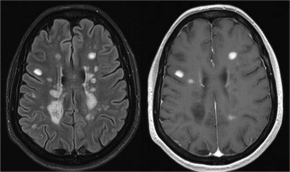
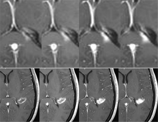
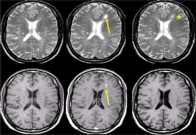
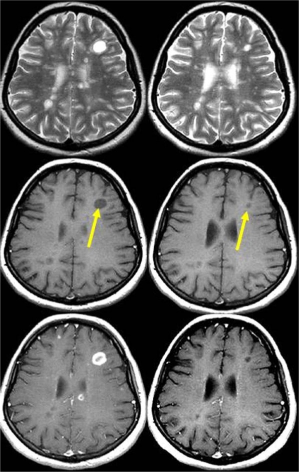
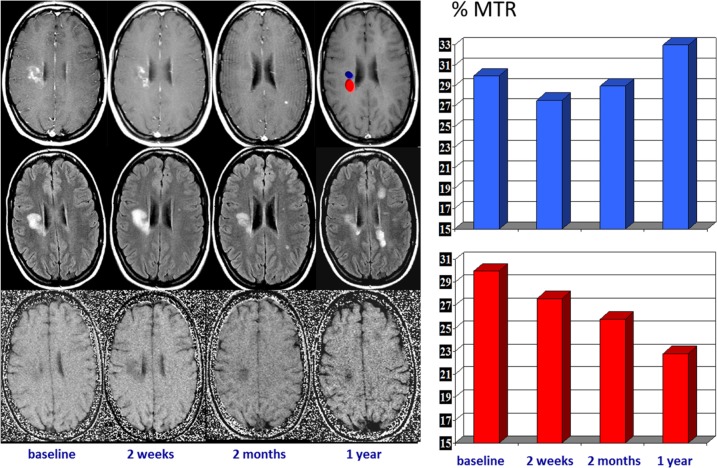
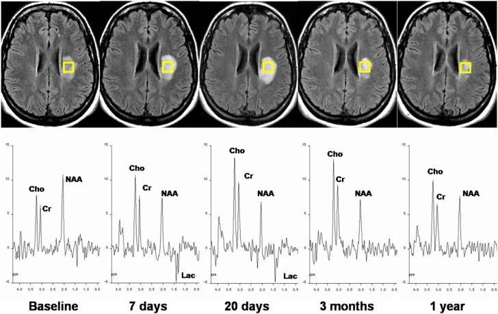
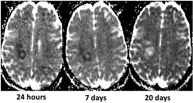
Disease activity in multiple sclerosis (MS) is strongly linked to the formation of new lesions, which involves a complex sequence of inflammatory, degenerative, and reparative processes. Conventional magnetic resonance imaging (MRI) techniques, such as T2-weighted and gadolinium-enhanced T1-weighted sequences, are highly sensitive in demonstrating the spatial and temporal dissemination of demyelinating plaques in the brain and spinal cord. Hence, these techniques can provide quantitative assessment of disease activity in patients with MS, and they are commonly used in monitoring treatment efficacy in clinical trials and in individual cases. However, the correlation between conventional MRI measures of disease activity and the clinical manifestations of the disease, particularly irreversible disability, is weak. This has been explained by a process of exhaustion of both structural and functional redundancies that increasingly prevents repair and recovery, and by the fact that these imaging techniques do not suffice to explain the entire spectrum of the disease process and lesion development. Nonconventional MRI techniques, such as magnetization transfer imaging, diffusion-weighted imaging, and proton magnetic resonance spectroscopy, which can selectively measure the more destructive aspects of MS pathology and monitor the reparative mechanisms of this disease, are increasingly being used for serial analysis of new lesion formation and provide a better approximation of the pathological substrate of MS plaques. These nonconventional MRI-based measures better assess the serial changes in newly forming lesions and improve our understanding of the relationship between the damaging and reparative mechanisms that occur in MS.

Keywords: diffusion-weighted imaging; lesion development; magnetic resonance imaging; magnetization transfer imaging; multiple sclerosis; proton magnetic resonance spectroscopy.


Conflict of interest statement

Conflict of interest statement: The authors declare no conflicts of interest in preparing this article.

Figures

Figure 1.
Figure 1.
Conventional magnetic resonance imaging in multiple sclerosis. T2-FLAIR (left) and gadolinium-enhanced T1-weighted (right) sequences. T2-FLAIR image shows multiple focal demyelinating lesions that are hyperintense relative to the normal appearing brain tissue. After contrast administration, some of the lesions are hyperintense on T1-weighted images, indicating increased permeability of the blood–brain barrier, a feature that distinguishes acute from chronic demyelinating lesions. FLAIR, fluid attenuation inversion recovery.
Figure 2.
Figure 2.
Evolution of contrast uptake in a newly formed lesion. Serial contrast-enhanced T1-weighted images obtained 5, 10, 15, and 20 min after gadolinium injection. A nodular-enhanced lesion located in the splenium of the corpus callosum (upper row) increases in size over time (centrifugal pattern), while an initial ring-enhanced lesion (lower row) becomes nodular (centripetal pattern).
Figure 3.
Figure 3.
Transverse T2-weighted (upper row) and contrast-enhanced T1-weighted (lower row) brain magnetic resonance imaging scans obtained serially at monthly intervals in a patient with multiple sclerosis. Observe formation of a new plaque in the left frontal white matter showing transient contrast uptake (arrow). With cessation of inflammatory activity, the T2 lesion decreased in size, but left a persistent hyperintense footprint on the T2-weighted image (asterisk).
Figure 4.
Figure 4.
Serial magnetic resonance imaging (MRI) scans obtained in a patient with relapsing–remitting multiple sclerosis. T2-weighted (upper row), unenhanced T1-weighted (middle row), and contrast-enhanced T1-weighted (lower row) MRI scans obtained at baseline (left) and 1 year later (right). Observe the active ‘black hole’ (nodular enhancement) in the subcortical white matter of the right frontal lobe (arrow), which becomes isointense on T1-weighted imaging with cessation of inflammatory activity (no enhancement).
Figure 5.
Figure 5.
Serial magnetic resonance imaging (MRI) scans obtained in a patient with relapsing–remitting multiple sclerosis. T2-weighted (upper row), unenhanced T1-weighted (middle row), and contrast-enhanced T1-weighted (lower row) MRI scans obtained at baseline (left) and 1 year later (right). Observe the active ‘black hole’ in the subcortical white matter of the left frontal lobe (arrow), which shows a ring-enhancement pattern of contrast uptake. After 1 year, the lesion decreased in size (arrow), but remained hypointense on T1-weighted images, indicating an irreversible black hole.
Figure 6.
Figure 6.
Serial magnetic resonance imaging changes occurring in an acute multiple sclerosis plaque located in the right centrum ovale. Contrast-enhanced T1-weighted images (upper row), T2-FLAIR images (middle row), and magnetization transfer (MTR) maps (lower row) obtained at baseline and at 7 days, 1 month, and 1 year later. The acute enhanced plaque shows the typical waxing and waning characteristics on T2-weighted images. The initial T2 lesion shrinkage observed after cessation of contrast uptake can be interpreted as a consequence of resorption of inflammatory edema, but the subsequent extended size decrease likely reflects a repair process. The serial MTR maps show two different components within the lesion, one with partial MTR recovery, likely reflecting remyelination, and the other with a progressive MTR decrease, likely reflecting ongoing demyelination (on the right, a plot of the serial MTR values obtained at the two locations). FLAIR, fluid attenuation inversion recovery.
Figure 7.
Figure 7.
Serial magnetic resonance imaging and spin-echo spectra recorded at an echo time of 135 ms from an acute multiple sclerosis plaque. T2-FLAIR images show an initial progressive lesion size increase followed by a decrease over 1 year of follow up. 1H-MRS during the acute stage shows the presence of Lac, a slight decrease in NAA, and an increase in Cho. The longitudinal study demonstrates Lac disappearance at 3 months, persistent low levels of NAA, a progressive Cho increase during the first weeks followed by partial recovery, and relatively stable Cr at all time points. 1H-MRS, proton magnetic resonance spectroscopy; Cho, choline; Cr, creatine; FLAIR, fluid attenuation inversion recovery; Lac, lactate; NAA, N-acetylaspartate.
Figure 8.
Figure 8.
Transient reduced diffusivity of an acute multiple sclerosis plaque (same lesion as in Figure 7) in a 22-year-old woman who was admitted to the hospital for sudden dysarthria, numbness of the right hand, and ensuing right hemiparesis. The apparent diffusion coefficient (ADC) maps obtained serially after onset of symptoms show an initially low ADC, likely reflecting cytotoxic edema or dense inflammatory cell infiltration, followed by a progressive ADC increase paralleling the development of vasogenic edema.

References

    1. Arnold D., De Stefano N., Narayanan S., Matthews P. (2000) Proton MR spectroscopy in multiple sclerosis. Neuroimaging Clin N Am 10: 789–798 - PubMed
    1. Arnold D., Matthews P., Francis G., O’Connor J., Antel J. (1992) Proton magnetic resonance spectroscopic imaging for metabolic characterization of demyelinating plaques. Ann Neurol 31: 235–241 - PubMed
    1. Barkhof F. (2002) The clinico-radiological paradox in multiple sclerosis revisited. Curr Opin Neurol 15: 239–245 - PubMed
    1. Barkhof F., Calabresi P., Miller D., Reingold S.(2009) Imaging outcomes for neuroprotection and repair in multiple sclerosis trials. Nat Rev Neurol 5: 256–266 - PubMed
    1. Budde M., Kim J., Liang H., Schmidt R., Russell J., Cross A., et al. (2007) Toward accurate diagnosis of white matter pathology using diffusion tensor imaging. Magn Reson Med 57: 688–695 - PubMed