Review
. 2013 Oct;445(1-2):232-43.
doi: 10.1016/j.virol.2013.07.018. Epub 2013 Aug 31.

Human papillomavirus genome variants

Affiliations
Review

Human papillomavirus genome variants

Robert D Burk et al. Virology. 2013 Oct.

Abstract

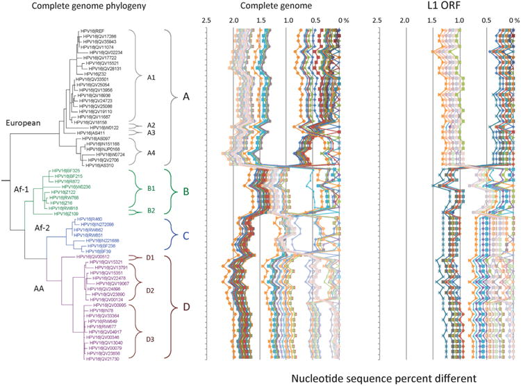
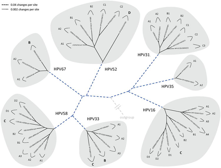
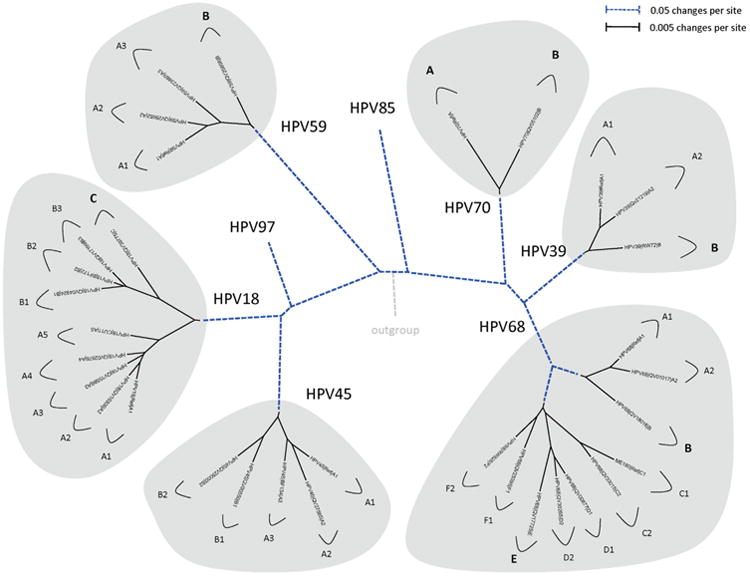
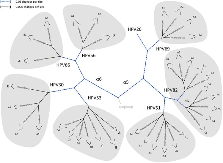
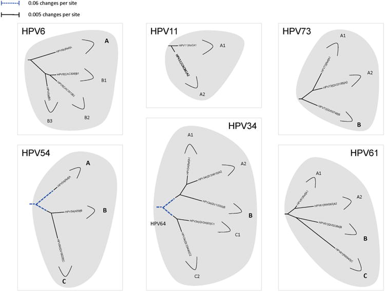
Amongst the human papillomaviruses (HPVs), the genus Alphapapillomavirus contains HPV types that are uniquely pathogenic. They can be classified into species and types based on genetic distances between viral genomes. Current circulating infectious HPVs constitute a set of viral genomes that have evolved with the rapid expansion of the human population. Viral variants were initially identified through restriction enzyme polymorphisms and more recently through sequence determination of viral fragments. Using partial sequence information, the history of variants, and the association of HPV variants with disease will be discussed with the main focus on the recent utilization of full genome sequence information for variant analyses. The use of multiple sequence alignments of complete viral genomes and phylogenetic analyses have begun to define variant lineages and sublineages using empirically defined differences of 1.0-10.0% and 0.5-1.0%, respectively. These studies provide the basis to define the genetics of HPV pathogenesis.

Keywords: Alphapapillomaviruses; HPV; HPV evolution; HPV variant lineages; Human papillomavirus variants.


Figures

Fig.1
Fig.1
HPV16 variant tree topology and pairwise comparisons of individual complete genomes. A maximum likelihood (ML) tree was inferred from a global alignment of 62 complete genome nucleotide sequences of HPV16 using RAxML HPC v7.2.8 (Stamatakis, 2006). The 62 HPV16 genomes were previously described in Smith et al. (2011). Distinct variant lineages (i.e., termed A/B/C/D) and sublineages (e.g., termed A1/A2/A3/A4) were classified according to the topology and nucleotide sequence differences from > 1% to < 10%, and > 0.5% to < 1% ranges, respectively (Bernard et al., 2010; Chen et al., 2011). The p-distance method in MEGA5 (Tamura et al., 2011) was used to calculate pairwise nucleotide sequence differences for each isolate compared to all other isolates based on the complete genome nucleotide sequences and are shown in the middle panel, labeled “Complete genome” at the top of the figure. For comparison, the L1 ORF nucleotide sequence of each isolate was compared to that of all other isolates and is shown in the right panel, labeled “L1 ORF”. Values for each comparison of a given isolate are connected by lines and the comparison to self is indicated by the 0% difference point.
Fig. 2
Fig. 2
Phylogenetic trees showing representative types and variant lineages/sublineages of alpha-9. A maximum likelihood tree was constructed using RAxML HPC v7.2.8 (Stamatakis, 2006) inferred from the global alignment of complete genome nucleotide sequences using the program MUSCLE (Edgar, 2004) linearized at the first ATG of the E1 ORF. Representative genomes of each lineage and/or sublineage are listed in Table 1, with GenBank accession numbers. The bold part represents groupings of each type. The length of dashed and solid lines represents distance between isolates, although the number of changes is different for these two lines; the scale is indicated in the upper left corner of Figs. 2, 4 and 5; it is shown in the upper right corner of Fig. 3.
Fig. 3
Fig. 3
Phylogenetic trees showing representative types and variant lineages/sublineages of alpha-7. A maximum likelihood tree was constructed using RAxML HPC v7.2.8 (Stamatakis, 2006) inferred from the global alignment of complete genome nucleotide sequences using the program MUSCLE (Edgar, 2004) linearized at the first ATG of the E1 ORF. Representative genomes of each lineage and/or sublineage are listed in Table 1, with GenBank accession numbers. The bold part represents groupings of each type. The length of dashed and solid lines represents distance between isolates, although the number of changes is different for these two lines; the scale is indicated in the upper left corner of Figs. 2, 4 and 5; it is shown in the upper right corner of Fig. 3.
Fig. 4
Fig. 4
Phylogenetic trees showing representative types and variant lineages/sublineages. A maximum likelihood tree was constructed using RAxML HPC v7.2.8 (Stamatakis, 2006) inferred from the global alignment of complete genome nucleotide sequences using the program MUSCLE (Edgar, 2004) linearized at the first ATG of the E1 ORF. Representative genomes of each lineage and/or sublineage are listed in Table 1, with GenBank accession numbers. The bold part represents groupings of each type. The length of dashed and solid lines represents distance between isolates, although the number of changes is different for these two lines; the scale is indicated in the upper left corner of Figs. 2, 4 and 5; it is shown in the upper right corner of Fig. 3.
Fig. 5
Fig. 5
Phylogenetic trees showing representative types and variant lineages/sublineages. A maximum likelihood tree was constructed using RAxML HPC v7.2.8 (Stamatakis, 2006) inferred from the global alignment of complete genome nucleotide sequences using the program MUSCLE (Edgar, 2004) linearized at the first ATG of the E1 ORF. Representative genomes of each lineage and/or sublineage are listed in Table 1, with GenBank accession numbers. The bold part represents groupings of each type. The length of dashed and solid lines represents distance between isolates, although the number of changes is different for these two lines; the scale is indicated in the upper left corner of Figs. 2, 4 and 5; it is shown in the upper right corner of Fig. 3.

Similar articles

Cited by

References

    1. Alizon S, Luciani F, Regoes RR. Epidemiological and clinical consequences of within-host evolution. Trends Microbiol. 2011;19:24–32. - PubMed
    1. Arias-Pulido H, Peyton CL, Torrez-Martinez N, Anderson DN, Wheeler CM. Human papillomavirus type 18 variant lineages in United States populations characterized by sequence analysis of LCR-E6, E2, and L1 regions. Virology. 2005;338:22–34. - PubMed
    1. Barzon L, Militello V, Lavezzo E, Franchin E, Peta E, Squarzon L, Trevisan M, Pagni S, Dal Bello F, Toppo S, Palu G. Human papillomavirus genotyping by 454 next generation sequencing technology. J Clin Virol. 2011;52:93–97. - PubMed
    1. Bennett MD, Reiss A, Stevens H, Heylen E, Van Ranst M, Wayne A, Slaven M, Mills JN, Warren KS, O'Hara AJ, Nicholls PK. The first complete papillomavirus genome characterized from a marsupial host: a novel isolate from Bettongia penicillata. J Virol. 2010;84:5448–5453. - PMC - PubMed
    1. Bernard HU. Coevolution of papillomaviruses with human populations. Trends Microbiol. 1994;2:140–143. - PubMed

Publication types

Substances