Review
. 2013 Sep;123(9):3664-71.
doi: 10.1172/JCI67230. Epub 2013 Sep 3.

HIF-1 mediates metabolic responses to intratumoral hypoxia and oncogenic mutations

Affiliations
Review

HIF-1 mediates metabolic responses to intratumoral hypoxia and oncogenic mutations

Gregg L Semenza. J Clin Invest. 2013 Sep.

Abstract

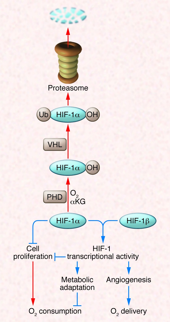
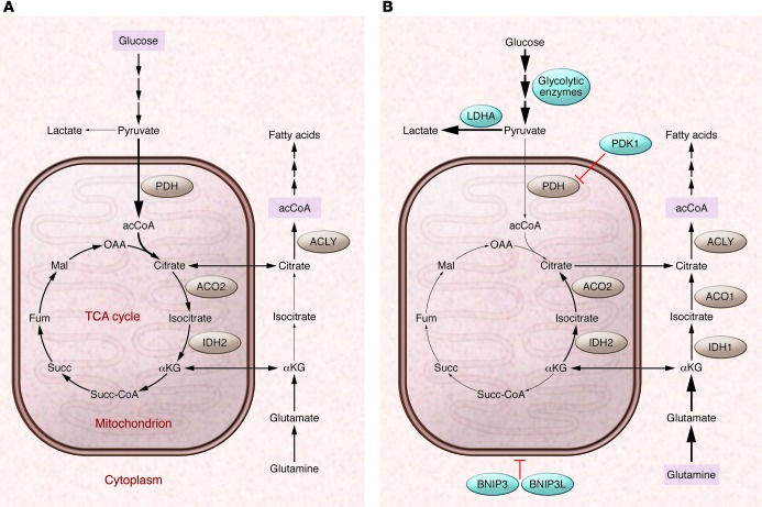
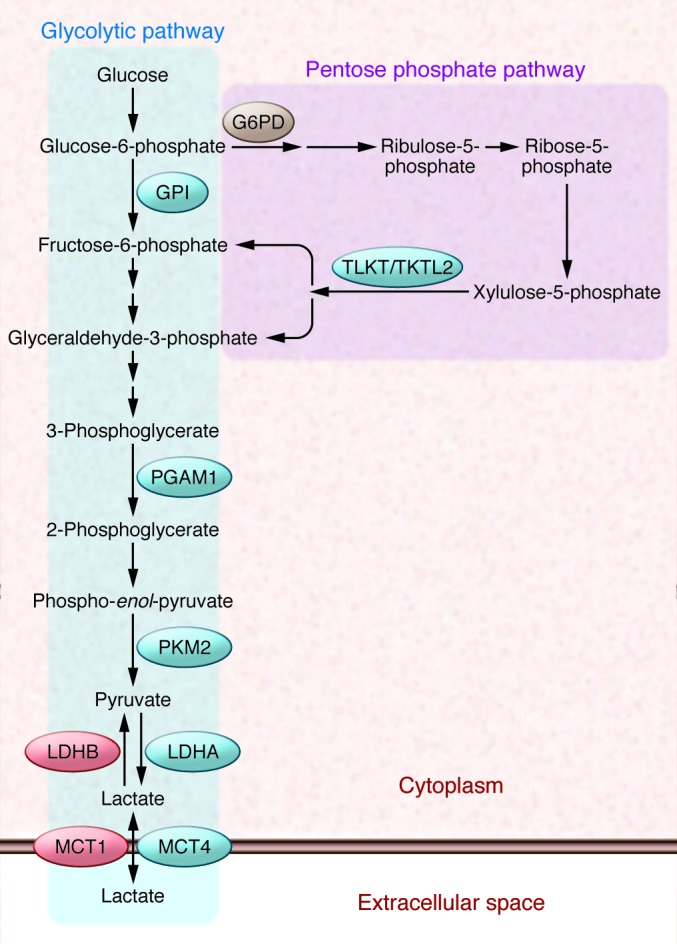
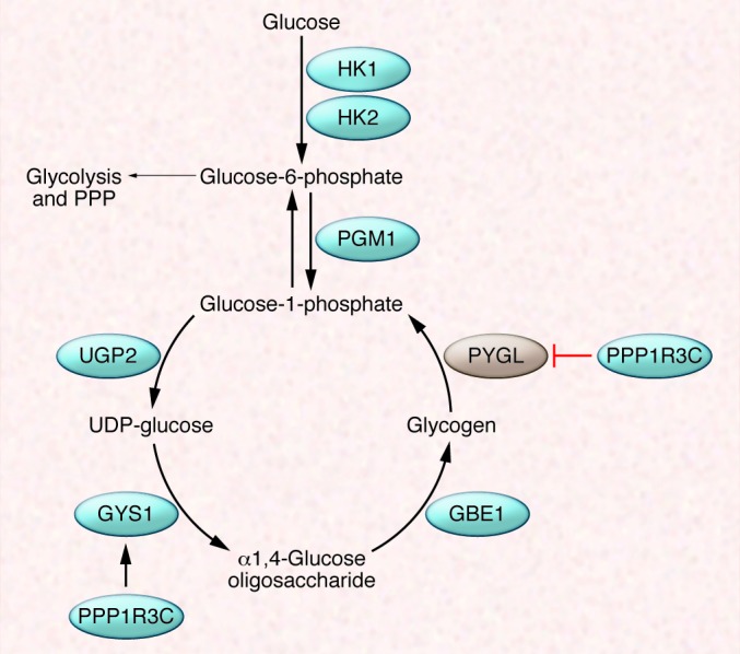
Hypoxia occurs frequently in human cancers and induces adaptive changes in cell metabolism that include a switch from oxidative phosphorylation to glycolysis, increased glycogen synthesis, and a switch from glucose to glutamine as the major substrate for fatty acid synthesis. This broad metabolic reprogramming is coordinated at the transcriptional level by HIF-1, which functions as a master regulator to balance oxygen supply and demand. HIF-1 is also activated in cancer cells by tumor suppressor (e.g., VHL) loss of function and oncogene gain of function (leading to PI3K/AKT/mTOR activity) and mediates metabolic alterations that drive cancer progression and resistance to therapy. Inhibitors of HIF-1 or metabolic enzymes may impair the metabolic flexibility of cancer cells and make them more sensitive to anticancer drugs.


Figures

Figure 1
Figure 1. HIF-1 regulates the balance between O2 supply and demand.
In well-oxygenated cells, prolyl hydroxylase domain (PHD) proteins use O2 and α-ketoglutarate (αKG) to hydroxylate HIF-1α, which is then bound by VHL, ubiquitylated, and degraded by the proteasome. Under hypoxic conditions, the hydroxylation reaction is inhibited and HIF-1α accumulates and regulates cell proliferation directly or dimerizes with HIF-1β to activate the transcription of hundreds of target genes, many of which encode enzymes and transporters that control cell metabolism. Red and blue arrows indicate reactions that are favored in aerobic and hypoxic conditions, respectively.
Figure 2
Figure 2. Oxygen-dependent regulation of glucose and glutamine metabolism by HIF-1.
(A) In well-oxygenated cells, glucose is metabolized to pyruvate, which is converted to acCoA by PDH for entry into the TCA cycle. Glucose-derived citrate is shuttled to the cytosol and converted to acCoA by ATP-citrate lyase (ACLY) for fatty acid synthesis. Glutamine is converted to αKG for entry into the TCA cycle. (B) In hypoxic cells, HIF-1 activates the transcription of several genes: (a) PDK1 and genes encoding other isoforms of PDH kinase, which phosphorylates and inactivates PDH; (b) LDHA, which converts pyruvate to lactate; (c) other glycolytic enzymes and glucose transporters to increase flux through the glycolytic pathway; and (d) BNIP3 and BNIP3L, which trigger mitochondrial selective autophagy. To compensate for the reduced flux of glucose to citrate, reductive carboxylation of glutamine generates cytosolic citrate for fatty acid synthesis. Note the change in direction of the reactions catalyzed by aconitase (ACO1, ACO2) and isocitrate dehydrogenase (IDH1, IDH2) under hypoxic conditions. Blue circles indicate proteins that are products of HIF-1 target genes. Fum, fumarate; Mal, malate; OAA, oxaloacetate; Succ, succinate; Succ-CoA, succinyl coenzyme A.
Figure 3
Figure 3. Glucose flux through the glycolytic (Embden-Meyerhof) pathway and pentose phosphate pathway (PPP).
G6PD shunts glucose-6-phosphate away from glycolysis and into the oxidative arm of the PPP. Transketolase (TKT) and transketolase-like 2 (TKTL2) shunt xylulose-5-phosphate back into the glycolytic pathway. Only those glycolytic enzymes that are discussed specifically in the text are shown: glucose phosphate isomerase (GPI), PGAM1, PKM2, and LDHA and LDHB. Also shown are MCT4 and MCT1, which transport lactate out of and into cells, respectively.
Figure 4
Figure 4. Glycogen synthesis and glycogenolysis.
PGM1 shunts glucose-6-phosphate away from glycolysis for use in glycogen synthesis, which requires the activity of three enzymes: UGP2, GYS1, and GBE1. Glucose-1-phosphate is released from glycogen (glycogenolysis) by PYGL. GYS1 and PYGL are positively and negatively regulated by protein phosphatase 1 regulatory subunit 3C (PPP1R3C).

References

    1. Wang GL, Semenza GL. Purification and characterization of hypoxia-inducible factor 1. J Biol Chem. 1995;270(3):1230–1237. doi: 10.1074/jbc.270.3.1230. - DOI - PubMed
    1. Wang GL, Jiang BH, Rue EA, Semenza GL. Hypoxia-inducible factor 1 is a basic-helix-loop-helix-PAS heterodimer regulated by cellular O2 tension. . Proc Natl Acad Sci U S A. 1995;92(12):5510–5514. doi: 10.1073/pnas.92.12.5510. - DOI - PMC - PubMed
    1. Semenza GL, et al. Hypoxia response elements in the aldolase A, enolase 1, and lactate dehydrogenase A gene promoters contain essential binding sites for hypoxia-inducible factor 1. J Biol Chem. 1996;271(51):32529–32537. doi: 10.1074/jbc.271.51.32529. - DOI - PubMed
    1. Kaelin WG, Jr, Ratcliffe PJ. Oxygen sensing by metazoans: the central role of the HIF hydroxylase pathway. Mol Cell. 2008;30(4):393–402. doi: 10.1016/j.molcel.2008.04.009. - DOI - PubMed
    1. Tian H, McKnight SL, Russell DW. Endothelial PAS domain protein 1 (EPAS1), a transcription factor selectively expressed in endothelial cells. Genes Dev. 1997;11(1):72–82. doi: 10.1101/gad.11.1.72. - DOI - PubMed

Publication types

Substances