. 2014 Oct;13(5):455-68.
doi: 10.7785/tcrtexpress.2013.600254. Epub 2013 Aug 31.

Large field, high resolution full-field optical coherence tomography: a pre-clinical study of human breast tissue and cancer assessment

Affiliations

Large field, high resolution full-field optical coherence tomography: a pre-clinical study of human breast tissue and cancer assessment

Osnath Assayag et al. Technol Cancer Res Treat. 2014 Oct.

Abstract

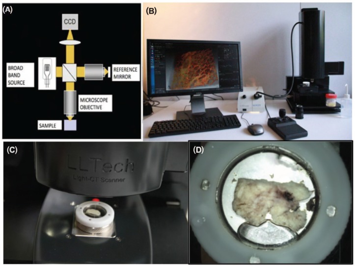
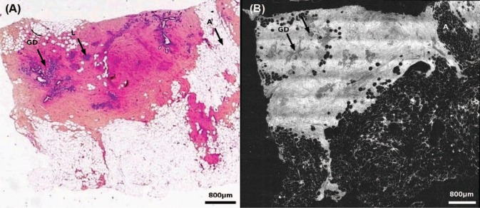
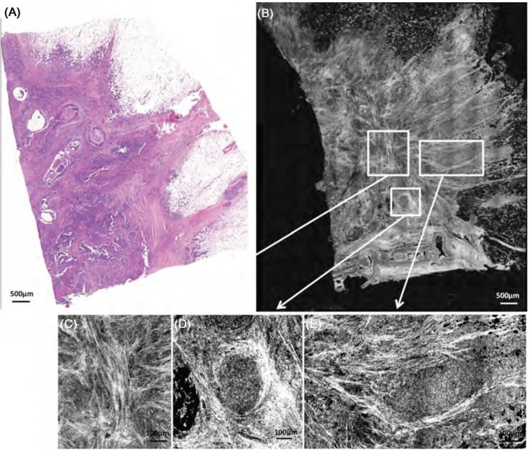
We present a benchmark pilot study in which high-resolution Full-Field Optical Coherence Tomography (FF-OCT) was used to image human breast tissue and is evaluated to assess its ability to aid the pathologist's management of intra-operative diagnoses. FF-OCT imaging safety was investigated and agreement between FF-OCT and routinely prepared histopathological images was evaluated. The compact setup used for this study provides 1 mm3 resolution and 200 mm imaging depth, and a 2.25 cm2 specimen is scanned in about 7 minutes. 75 breast specimens were imaged from 22 patients (21 women, 1 man) with a mean age of 58 (range: 25-83). Pathologists blind diagnosed normal/benign or malignant tissue based on FF-OCT images alone, diagnosis from histopathology followed for comparison. The contrast in the FF-OCT images is generated by intrinsic tissue scattering properties, meaning that no tissue staining or preparation is required. Major architectural features and tissue structures of benign breast tissue, including adipocytes, fibrous stroma, lobules and ducts were characterized. Subsequently, features resulting from pathological modification were characterized and a diagnosis decision tree was developed. Using FF-OCT images, two breast pathologists were able to distinguish normal/benign tissue from lesional with a sensitivity of 94% and 90%, and specificity of 75% and 79% respectively.


Conflict of interest statement

Conflict of Interest: ACB and BdP have indirect stock ownership in LLTech SAS. LLTech SAS has not sponsored the study.

Figures

Figure 1:
Figure 1:
FF-OCT experimental setup: schematic of FF-OCT setup based on a Linnik interferometer configuration (A), photograph of compact set-up used for the study (B), the sample moves under the objective for imaging (C), the sample mounted in its holder (D).
Figure 2:
Figure 2:
Breast tissue basic structures. Lobule (A), galactophorous duct (B), cross section of a galactophorous duct with calcifications (C), vessel (D), adipocytes (E), scar fibrous tissue (F), normal fibrous tissue (G), fibrous tissue surrounding carcinomatous cells in tumourous stroma (H). FF-OCT images were acquired 20 µm beneath the tissue surface.
Figure 3:
Figure 3:
Diagnosis decision tree for FF-OCT images of human breast tissue.
Figure 4:
Figure 4:
Healthy breast tissue specimen. Post-menopausal woman. FF-OCT image was acquired 20 µm beneath the tissue surface. Whole slide image of the conventional gold-standard histology preparation (A) and the corresponding FF-OCT image (B). Characteristic structures of breast tissue such as the lobules (L), galactophorous ducts (GD), adipocytes (A) and a normal fibrous tissue are seen. Some entanglement of the fibrous tissue and adipose tissue is visible. The fibrous tissue appears grainy with a medium back scattering level. Ducts are cut longitudinally; thus they appear dark grey. A digital zooming on the image reveals acini in the lobules.
Figure 5:
Figure 5:
Different types of invasive adenocarcinoma. Stellate (A-D) and nodular (E-H). FF-OCT images were acquired 20 µm beneath the tissue surface. The trabeculae of the highly scattering tumor-associated fibrous tissue can be observed (F). This is in contrast to the normal fibrous tissue that appears grainy and produces less scattering (G). Furthermore, tumor-associated adipocytes are smaller than those outside the tumor (D). In the stellate tumor (B), the fibrous tissue is seen invading the adipose tissue. In the nodular tumor (F), foci of carcinoma cells appear as grey zones surrounded by the highly scattering tumorassociated fibrous tissue trabeculae (H). A circular dilated duct with secretion in the lumen is visible in the center of the nodule. The different aspect of fibrous tissue defines the tumor margins (G).
Figure 6:
Figure 6:
Ductal invasive adenocarcinoma with Ductal Carcinoma In Situ (DCIS) component. FF-OCT images were acquired 20 µm beneath the tissue surface. Histology (A) and FF-OCT image (B) with digital zooms (C-E). Enlarged lobules and ducts filled with DCIS (D, E) are clearly visible on the image. In addition, the invasive component is characterized by the presence of highly scattering fibrous tissue (C) and foci of darker grey carcinoma cells (E).
Figure 7:
Figure 7:
Ductal carcinoma. Histology (A, E) and FF-OCT (B-D, F, G) of a ductal carcinoma in situ (B) with an abnormal duct (C) and an enlarged lobule (D). Fibroadenoma (F) and an enlarged ductule (G). FF-OCT images were acquired 20 µm beneath the tissue surface. DCIS is noted in (B), characterized by enlarged abnormal lobules (D) and ducts (C), which can be distinguished easily in a digital zoom-in on the image. The acini in the enlarged lobule are clearly visible and the narrow lumen in the duct is indicative of malignancy.

Similar articles

Cited by

References

    1. American Cancer Society: Cancer Facts and Figures; 2011.
    1. Silverstein MJ, Recht A, Lagios MD, Bleiweiss IJ, Blumencranz PW, Gizienski T, Harms SE, Harness J, Jackman RJ, Klimberg VS, Kuske R, Levine GM, Linver MN, Rafferty EA, Rugo H, Schilling K, Tripathy D, Whitworth PW, Willey SC. Image-detected breast cancer: state-of-the-art diagnosis and treatment. J Am Coll Surg 209, 504–520 (2009). DOI: 10.1016/j.jamcollsurg.2009.07.006. - PubMed
    1. Cendán JC, Coco D, Copeland EM. Accuracy of intraoperative frozen-section analysis of breast cancer lumpectomy-bed margins. J Am Coll of Surg 201, 194–198 (2005). DOI: 10.1016/j.jamcollsurg.2005.03.014. - PubMed
    1. Nakazawa H, Rosen P, Lane N, Lattes R. Frozen section experience in 3000 cases: accuracy, limitations, and value in residence training. Am J Clin Pathol 49, 41–51 (1968). - PubMed
    1. Olson TP, Harter J, Munoz A, Mahvi DM, Breslin T. Frozen section analysis for intraoperative margin assessment during breast-conserving surgery results in low rates of re-excision and local recurrence. Ann Surg Oncol 14, 2953–2960 (2007). DOI: 10.1245/s10434-007-9437-1. - PubMed

Publication types