

A computational model of fibroblast and macrophage spatial/temporal dynamics in foreign body reactions
- PMID: 24001881
- PMCID: PMC3964857
- DOI: 10.1016/j.jim.2013.08.013
A computational model of fibroblast and macrophage spatial/temporal dynamics in foreign body reactions
Abstract
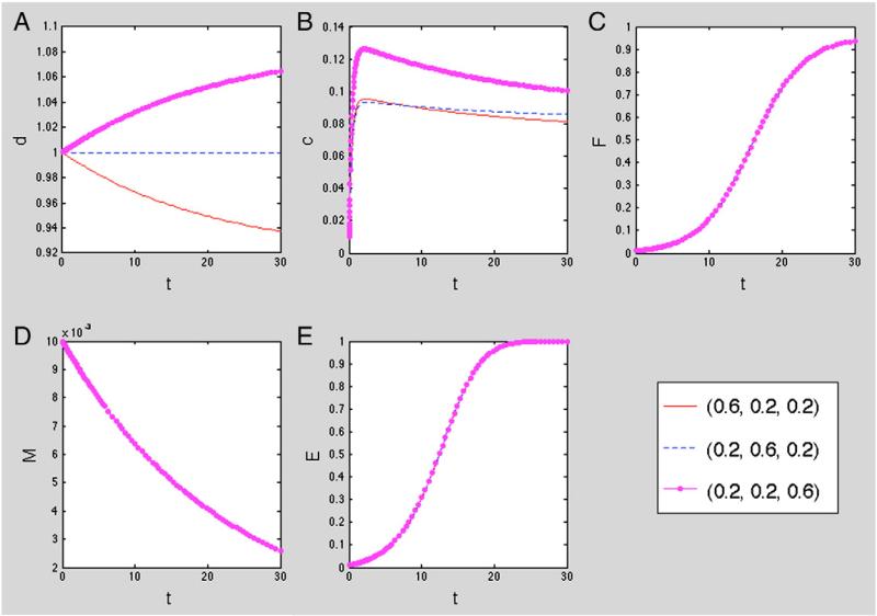
The implantation of medical devices often triggers several immune responses, one kind of which is categorized as foreign body reactions. It is well established that macrophages and many other cells participate in the complex processes of foreign body reactions, and cause severe inflammations and fibrotic capsule formation in surrounding tissues. However, the detailed mechanisms of macrophage responses, recruitment and activation, in foreign body reactions are not totally understood. In the meantime, mathematical models have been proposed to systematically decipher the behavior of this complex system of multiple cells, proteins and biochemical processes in wound healing responses. Based on these early works, this study introduces a mathematical model in two spatial dimensions to investigate the transient behavior of macrophages, fibroblasts and their interactions during the formation of fibrotic tissue. We find that the simulation results are consistent with the experimental observations. These findings support that the model can reveal quantitative insights for studying foreign body reaction processes.
Keywords: Computational model; Fibroblast; Foreign body reactions; Macrophage; Medical implant; Spatial and time dynamics.
© 2013. Published by Elsevier B.V. All rights reserved.
Figures
Similar articles
-
Stability analysis of a model for foreign body fibrotic reactions.Comput Math Methods Med. 2012;2012:809864. doi: 10.1155/2012/809864. Epub 2012 Sep 13. Comput Math Methods Med. 2012. PMID: 23193430 Free PMC article.
-
In vitro chemotaxis and tissue remodeling assays quantitatively characterize foreign body reaction.ALTEX. 2017;34(2):253-266. doi: 10.14573/altex.1610071. Epub 2016 Oct 11. ALTEX. 2017. PMID: 27725990
-
A mathematical model for foreign body reactions in 2D.Int J Comput Math. 2011 Feb;88(3):610-633. doi: 10.1080/00207161003640035. Int J Comput Math. 2011. PMID: 21532988 Free PMC article.
-
Beyond Encapsulation: Exploring Macrophage-Fibroblast Cross Talk in Implant-Induced Fibrosis.Tissue Eng Part B Rev. 2024 Dec;30(6):596-606. doi: 10.1089/ten.TEB.2023.0300. Epub 2024 Mar 27. Tissue Eng Part B Rev. 2024. PMID: 38420650 Review.
-
Foreign Body Reaction to Subcutaneous Implants.Adv Exp Med Biol. 2015;865:93-108. doi: 10.1007/978-3-319-18603-0_6. Adv Exp Med Biol. 2015. PMID: 26306445 Review.
Cited by
-
M1 Macrophages Are Predominantly Recruited to the Major Pelvic Ganglion of the Rat Following Cavernous Nerve Injury.J Sex Med. 2017 Feb;14(2):187-195. doi: 10.1016/j.jsxm.2016.12.012. J Sex Med. 2017. PMID: 28161077 Free PMC article.
-
Interaction of Ceramic Implant Materials with Immune System.Int J Mol Sci. 2023 Feb 20;24(4):4200. doi: 10.3390/ijms24044200. Int J Mol Sci. 2023. PMID: 36835610 Free PMC article. Review.
-
Towards predicting implant-induced fibrosis: A standardized network model of macrophage-fibroblast interactions.Comput Struct Biotechnol J. 2025 Jul 13;27:3251-3263. doi: 10.1016/j.csbj.2025.07.022. eCollection 2025. Comput Struct Biotechnol J. 2025. PMID: 40746411 Free PMC article.
-
Computational modeling of phagocyte transmigration for foreign body responses to subcutaneous biomaterial implants in mice.BMC Bioinformatics. 2016 Feb 29;17:111. doi: 10.1186/s12859-016-0947-3. BMC Bioinformatics. 2016. PMID: 26927968 Free PMC article.
References
-
- Anderson JM. Multinucleated giant cells. Curr. Opin. Hematol. 2000;7:40. - PubMed
-
- Appling WD, O'Brien WR, Johnston DA, Duvic M. Synergistic enhancement of type I and III collagen production in cultured fibroblasts by transforming growth factor-beta and ascorbate. FEBS Lett. 1989;250:541. - PubMed
-
- Blankson J, Persaud D, Siliciano RF. Latent reservoirs for HIV-1. Curr. Opin. Infect. Dis. 1999;12:5. - PubMed
Publication types
MeSH terms
Grants and funding
LinkOut - more resources
Full Text Sources
Other Literature Sources
