

Osteocyte-driven bone remodeling
- PMID: 24002178
- PMCID: PMC3947228
- DOI: 10.1007/s00223-013-9774-y
Osteocyte-driven bone remodeling
Abstract
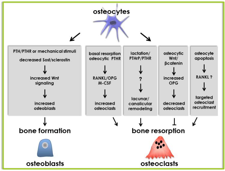
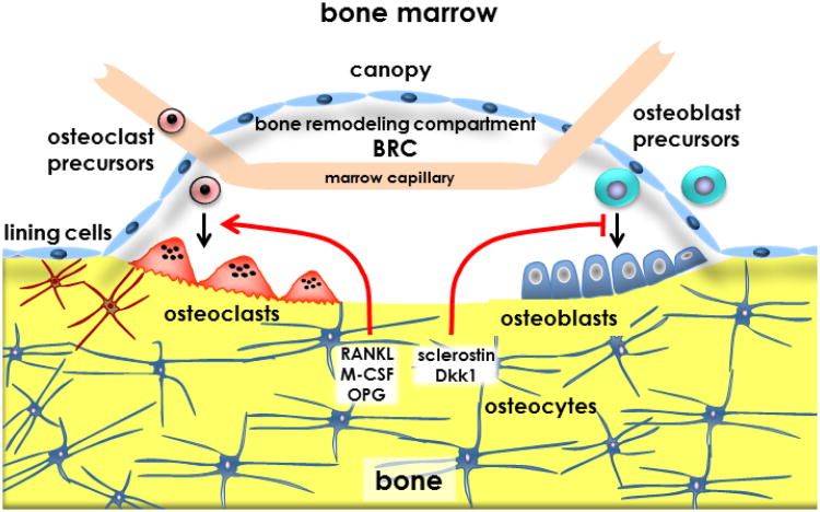
Osteocytes, the most abundant cells in bone, have been long postulated to detect and respond to mechanical and hormonal stimuli and to coordinate the function of osteoblasts and osteoclasts. The discovery that the inhibitor of bone formation sclerostin is primarily expressed in osteocytes in bone and downregulated by anabolic stimuli provided a mechanism by which osteocytes influence the activity of osteoblasts. Advances of the last few years provided experimental evidence demonstrating that osteocytes also participate in the recruitment of osteoclasts and the initiation of bone remodeling. Apoptotic osteocytes trigger yet-to-be-identified signals that attract osteoclast precursors to specific areas of bone, which in turn differentiate to mature, bone-resorbing osteoclasts. Osteocytes are also the source of molecules that regulate the generation and activity of osteoclasts, such as OPG and RANKL; and genetic manipulations of the mouse genome leading to loss or gain of function or to altered expression of either molecule in osteocytes markedly affect bone resorption. This review highlights these investigations and discusses how the novel concept of osteocyte-driven bone resorption and formation impacts our understanding of the mechanisms by which current therapies control bone remodeling.
Conflict of interest statement
The author has stated that there is no conflict of interest.
Figures
Similar articles
-
The osteocyte as a signaling cell.Physiol Rev. 2022 Jan 1;102(1):379-410. doi: 10.1152/physrev.00043.2020. Epub 2021 Aug 2. Physiol Rev. 2022. PMID: 34337974 Free PMC article. Review.
-
Osteocyte control of bone remodeling: is sclerostin a key molecular coordinator of the balanced bone resorption-formation cycles?Osteoporos Int. 2014 Dec;25(12):2685-700. doi: 10.1007/s00198-014-2808-0. Epub 2014 Jul 17. Osteoporos Int. 2014. PMID: 25030653 Review.
-
Regulatory mechanisms of RANKL presentation to osteoclast precursors.Curr Osteoporos Rep. 2014 Mar;12(1):115-20. doi: 10.1007/s11914-014-0189-0. Curr Osteoporos Rep. 2014. PMID: 24477414 Review.
-
The role of osteocytes in targeted bone remodeling: a mathematical model.PLoS One. 2013 May 22;8(5):e63884. doi: 10.1371/journal.pone.0063884. Print 2013. PLoS One. 2013. PMID: 23717504 Free PMC article.
-
Effects of PTH on osteocyte function.Bone. 2013 Jun;54(2):250-7. doi: 10.1016/j.bone.2012.09.016. Epub 2012 Sep 24. Bone. 2013. PMID: 23017659 Free PMC article. Review.
Cited by
-
HIF-1α activation impairs dendrites formation and elongation in osteocytogenesis.Heliyon. 2024 Jun 17;10(12):e32889. doi: 10.1016/j.heliyon.2024.e32889. eCollection 2024 Jun 30. Heliyon. 2024. PMID: 39005918 Free PMC article.
-
Maintaining homeostatic control of periodontal bone tissue.Periodontol 2000. 2021 Jun;86(1):157-187. doi: 10.1111/prd.12368. Epub 2021 Mar 10. Periodontol 2000. 2021. PMID: 33690918 Free PMC article. Review.
-
Osteocyte Remodeling of the Lacunar-Canalicular System: What's in a Name?Curr Osteoporos Rep. 2023 Feb;21(1):11-20. doi: 10.1007/s11914-022-00766-3. Epub 2022 Dec 13. Curr Osteoporos Rep. 2023. PMID: 36512204 Free PMC article. Review.
-
Osteocyte death during orthodontic tooth movement in mice.Angle Orthod. 2014 Nov;84(6):1086-92. doi: 10.2319/110713-813.1. Epub 2014 Apr 2. Angle Orthod. 2014. PMID: 24694014 Free PMC article.
-
Friend or Foe? Essential Roles of Osteoclast in Maintaining Skeletal Health.Biomed Res Int. 2020 Mar 3;2020:4791786. doi: 10.1155/2020/4791786. eCollection 2020. Biomed Res Int. 2020. PMID: 32190665 Free PMC article. Review.
References
-
- Holmbeck K, Bianco P, Caterina J, Yamada S, Kromer M, Kuznetsov SA, Mankani M, et al. MT1-MMP-deficient mice develop dwarfism, osteopenia, arthritis, and connective tissue disease due to inadequate collagen turnover. Cell. 1999;99:81–92. - PubMed
-
- Holmbeck K, Bianco P, Pidoux I, Inoue S, Billinghurst RC, Wu W, Chrysovergis K, et al. The metalloproteinase MT1-MMP is required for normal development and maintenance of osteocyte processes in bone. J Cell Sci. 2005;118:147–156. - PubMed
-
- Boyce BF, Xing L, Jilka RL, Bellido T, Weinstein RS, Parfitt AM, Manolagas SC. Apoptosis in bone cells. In: Bilezikian JP, Raisz LG, Rodan GA, editors. Principles of Bone Biology. Academic Press; San Diego, San Francisco, New York, London, Sydney, Tokyo: 2002. pp. 151–168.
Publication types
MeSH terms
Grants and funding
LinkOut - more resources
Full Text Sources
Other Literature Sources
