. 2013 Dec;115(3):343-51.
doi: 10.1007/s11060-013-1235-y. Epub 2013 Sep 4.

Heparin blocks transfer of extracellular vesicles between donor and recipient cells

Affiliations

Heparin blocks transfer of extracellular vesicles between donor and recipient cells

Nadia A Atai et al. J Neurooncol. 2013 Dec.

Abstract

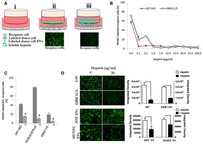
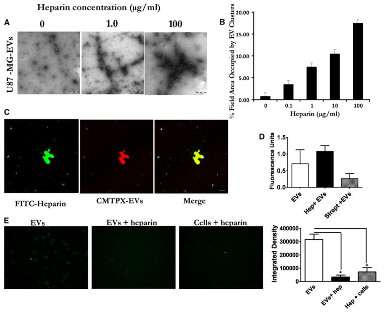
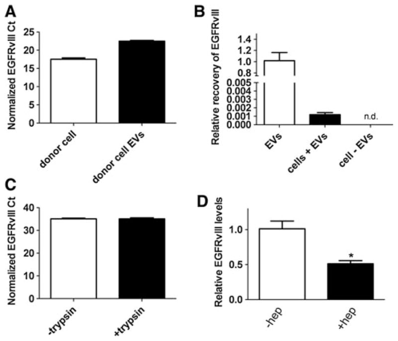
Extracellular vesicles (EVs) have been implicated in tumorigenesis. Biomolecules which can block EV binding and uptake into recipient cells may be of therapeutic value as well as enhance understanding of EV biology. Here, we show that heparin interacts with uptake of tumor-derived as well as non-tumor-derived EVs into recipient cells. Incubation of glioma cell-derived EVs with heparin resulted in micron-sized structures observed by transmission electron microscopy, with EVs clearly visible within these structures. Inclusion of heparin greatly diminished transfer of labeled EVs from donor to recipient tumor cells. We also show a direct interaction between heparin and EVs using confocal microscopy. We found that the block in EV uptake was at the level of cell binding and not internalization. Finally, incubation of glioma-derived EVs containing EGFRvIII mRNA with heparin reduced transfer of this message to recipient cells. The effect of heparin on EVs uptake may provide a unique tool to study EV function. It may also foster research of heparin or its derivatives as a therapeutic for disease in which EVs play a role.


Figures

Fig. 1
Fig. 1
Heparin blocks extracellular vesicle (EV) uptake in recipient cells. a Schematic of experiment of recipient cells uptake of donor cell EVs. (i). Recipient cells are plated in a well. PKH67-labeled donor cells are then added in the upper chamber of a transwell system in the absence (ii) or presence of heparin (iii). 48 h later, recipient cells are then examined for PKH67-labeled EV uptake by fluorescence microscopy (fluorescence images) and flow cytometry. b Flow cytometric quantitation of PKH67-labeled D384 donor cell-derived EVs uptake by recipient glioma cells in the presence of various concentrations of heparin. c Flow cytometric detection of PKH67-labeled U87-MG, Gli36-EGFRvIII, and GBM11/5 donor cell-derived EV uptake in presence or absence of 20 μg/ml heparin into unlabeled U87-MG, Gli36-EGFRvIII and GBM11/5 recipient cells, respectively. d Uptake of purified PKH67-labeled U87 and GBM 11/5-derived EVs into their respective unlabeled recipients (top panels and graph) or 293T-derived or HUVEC-derived EVs into recipient unlabeled HUVEC cells (bottom panels and graph) in presence or absence of 20 μg/ml heparin. *p <0.05
Fig. 2
Fig. 2
Heparin causes EV aggregation and binds EVs. a Suspensions of EVs derived from U87-MG cells in PBS were incubated with 0–100 μg/ml of heparin for 30 min at RT and then imaged by transmission EM (TEM). Scale bar = 1 μm. b Quantification of the TEM images show increased area occupied by the EV clusters as heparin concentration increases. (C) Confocal imaging of complexed FITC-heparin (green) and U87-MG -derived EVs (CMTPX-red). Merging of the images of FITC-heparin and EVs shows colocalization of heparin and EVs in yellow (Scale bar = 10 μm). d Incubating heparin with EVs increases their pelleting efficiency. PKH67 labeled U87 EVs were mixed with PBS, heparin, or streptavidin and pelleted 2 h at 100,000×g. Pelleted EVs were resuspended and fluorescence activity measured using a plate reader. e Binding heparin to cells reduces EV uptake. U87 glioma cells were incubated on ice with PBS or heparin (200 μg/ml) before adding PKH67 labeled U87-derived EVs. For control we incubated heparin and EVs at room temperature (EVs + heparin) before adding to cells. Magnification × 20
Fig. 3
Fig. 3
Heparin partially blocks oncogenic EGFRvIII mRNA transfer. a Relative levels of EGFRvIII mRNA in Gli36-EGFRvIII donor cells and their EVs. RNA was extracted from cells or EVs and 1 μg of RNA was used as template for a cDNA reaction. EGFRvIII Ct values were normalized to GAPDH Ct values for each sample. b Transfer of EGFRvIII mRNA inside EVs to recipient U87-MG cells (which lack endogenous EGFRvIII). 3.3 × 10⁹ Gli36-EGFRvIII derived EVs were added to cells and after 3 h at 37 °C RNA was isolated and a RT-qPCR performed to detect EGFRvIII mRNA. After normalization to GAPDH the levels of EGFRvIII were compared to the input levels from donor cell EVs. n.d. = not detectable. c Detected EGFRvIII message is on the inside of recipient cells. After incubation for 3 h at 37 °C, cells were washed and then incubated with trypsin to remove any EVs bound to the cell surface. Control samples were treated with PBS. RNA was isolated and EGFRvIII cDNA detected with RT-qPCR. d Gli36-EGFRvIII derived EVs were incubated with or without heparin (100 μg/ml) for 30 min at RT and next were added directly to recipient U87-MG cells. After 3 h at 37 °C, the total RNA of recipient cells was extracted and used for detection of EGFRvIII mRNA with RT-qPCR. Values were normalized to GAPDH and then compared to the levels in the absence of heparin which was arbitrarily set to 1.0. The depicted graph is representative of one of four independent experiments

References

    1. Dolecek TA, Propp JM, Stroup NE, Kruchko C. CBTRUS statistical report: primary brain and central nervous system tumors diagnosed in the United States in 2005–2009. Neuro-Oncol. 2012;14 (suppl 5):v1–v49. - PMC - PubMed
    1. Al-Nedawi K, Meehan B, Rak J. Microvesicles: messengers and mediators of tumor progression. Cell Cycle. 2009;8(13):2014–2018. - PubMed
    1. Théry C, Zitvogel L, Amigorena S. Exosomes: composition, biogenesis and function. Nat Rev Immunol. 2002;2(8):569–579. - PubMed
    1. Chaput N, Taieb J, Andre F, Zitvogel L. The potential of exosomes in immunotherapy. Expert Opin Biol Ther. 2005;5:737–747. - PubMed
    1. Wieckowski E, Whiteside TL. Human tumor-derived vs dendritic cell-derived exosomes have distinct biologic roles and molecular profiles. Immunol Res. 2006;36(1–3):247–254. - PubMed

Publication types

MeSH terms