. 2013 Oct;27(10):1666-77.
doi: 10.1210/me.2013-1211. Epub 2013 Sep 3.

Persistently altered epigenetic marks in the mouse uterus after neonatal estrogen exposure

Affiliations

Persistently altered epigenetic marks in the mouse uterus after neonatal estrogen exposure

Wendy N Jefferson et al. Mol Endocrinol. 2013 Oct.

Abstract

Neonatal exposure to diethylstilbestrol (DES) causes permanent alterations in female reproductive tract gene expression, infertility, and uterine cancer in mice. To determine whether epigenetic mechanisms could explain these phenotypes, we first tested whether DES altered uterine expression of chromatin-modifying proteins. DES treatment significantly reduced expression of methylcytosine dioxygenase TET oncogene family, member 1 (TET1) on postnatal day 5; this decrease was correlated with a subtle decrease in DNA 5-hydroxymethylcytosine in adults. There were also significant reductions in histone methyltransferase enhancer of zeste homolog 2 (EZH2), histone lysine acetyltransferase 2A (KAT2A), and histone deacetylases HDAC1, HDAC2, and HDAC3. Uterine chromatin immunoprecipitation was used to analyze the locus-specific association of modified histones with 2 genes, lactoferrin (Ltf) and sine oculis homeobox 1 (Six1), which are permanently upregulated in adults after neonatal DES treatment. Three histone modifications associated with active transcription, histone H3 lysine 9 acetylation (H3K9ac), H3 lysine 4 trimethylation (H3K4me3), and H4 lysine 5 acetylation (H4K5ac) were enriched at specific Ltf promoter regions after DES treatment, but this enrichment was not maintained in adults. H3K9ac, H4K5ac, and H3K4me3 were enriched at Six1 exon 1 immediately after neonatal DES treatment. As adults, DES-treated mice had greater differences in H4K5ac and H3K4me3 occupancy at Six1 exon 1 and new differences in these histone marks at an upstream region. These findings indicate that neonatal DES exposure temporarily alters expression of multiple chromatin-modifying proteins and persistently alters epigenetic marks in the adult uterus at the Six1 locus, suggesting a mechanism for developmental exposures leading to altered reproductive function and increased cancer risk.


Figures
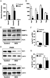
Figure 1.
Figure 1.
DES-induced alterations in uterine DNA methylation regulatory enzymes and DNA methylation. A, Relative mRNA expression of DNA methyltransferases on PND5. B, Relative mRNA expression of TET methylcytosine dioxygenases and thymine deglycosylase on PND5. C, Immunoblots of DNMT1 and DNMT3A on PND5. Actin was used as a loading control. In this and all subsequent immunoblots, each lane contains protein extract from a single animal, and n = 2–4 animals per group on each blot; protein from at least 4 animals was run in all cases. Representative blots are shown. D and E, Immunoblots of TET1 in PND5 (D) or adult ovx (E) uteri. Lamin was used as a loading control. F, Total DNA 5-methylcytosine. G, Total DNA 5hmC. For all graphs, mean ± SE is plotted; n = 4–8 per group. *, P < .05 compared with control at same time point.
Figure 2.
Figure 2.
DES-induced alterations in histone methyltransferases but not methylated histones on PND5. A, Relative mRNA expression of histone H3 methyltransferases on PND5. A–C, Genes encoding methyltransferases with major reported activity on H3K9 (A), H3K27 (B), and H3K4 (C). Mean ± SE is plotted; n = 4 per group. *, P < .05 compared with control. D, Immunoblot of EZH2. Actin was used as a loading control. E, Immunoblots of H3K9me2, H3K27me3, and H4K4me3. In this and all subsequent histone immunoblots, each lane contains 2 μg histones isolated from 2 uteri per sample, and n = 4 samples per group on each blot. In the bottom panel, total histones were stained on a duplicate gel to document equal protein loading.
Figure 3.
Figure 3.
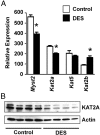
DES-induced alterations in histone-acetylating enzymes on PND5. A, Relative mRNA expression of histone acetyltransferases on PND5. Mean ± SE is plotted; n = 4 per group. *, P < .05 compared with control. B, Immunoblot of KAT2A in PND5 uteri. Actin was used as a loading control.
Figure 4.
Figure 4.
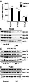
DES-induced alterations in HDACs but not acetylated histones. A, Relative mRNA expression of HDACs on PND5. Mean ± SE is plotted; n = 4 per group. *, P < .05 compared with control. B and C, Immunoblots of HDAC1, HDAC2, and HDAC3 in PND5 (B) or adult ovx (C) uteri. Actin was used as a loading control. D, Immunoblots of modified histones H3K9ac and H4K5ac. Total histone proteins were stained on a duplicate gel to ensure equal loading.
Figure 5.
Figure 5.
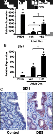
Permanent overexpression of Ltf and Six1 after neonatal DES treatment. A, Relative mRNA expression of the indicated gene in uteri from PND5, adult ovx vehicle (veh)-treated, or adult ovx plus estradiol (E2)-treated mice. Mean ± SE is plotted; n = 4 per group. *, P < .05 compared with control; †, P < .05 compared with the group indicated by the associated line. C, Immunohistochemical localization of SIX1 in the adult mouse uterus. Scale bars, 50 μm.
Figure 6.
Figure 6.
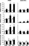
Differential association of modified histones with the Ltf gene region after neonatal DES exposure. ChIP assays were performed on uterine tissue from PND5 (left panels) or adult ovx (right panels) mice using the indicated modified histone antibodies. PCR was performed on a distal region and 2 regions near the Ltf transcription start site (CpG and ERE) (see Table 2 for primer locations). Mean ± SE is plotted; n = 4 per group. White bars represent control; black bars represent DES. *, P < .05 compared with control.
Figure 7.
Figure 7.
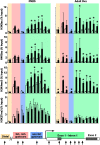
Persistent differential association of modified histones with the Six1 locus after neonatal DES exposure. ChIP assays were performed on uteri from PND5 (left panels) and adult ovx (right panels) mice using the indicated modified histone antibodies. PCR was performed on a distal region (tan), a GC-rich upstream region (pink), a non–GC-rich upstream region (blue), and a region covering exon 1 to intron 1 (green) of the Six1 gene (see Table 2 for primer locations). Mean ± SE is plotted; n = 4 per group. White bars represent control; black bars represent DES. *, P < .05 compared with control. A schematic representation of the Six1 gene indicating the primer locations is shown for reference.

References

    1. Schug TT, Janesick A, Blumberg B, Heindel JJ. Endocrine disrupting chemicals and disease susceptibility. J Steroid Biochem Mol Biol. 2011;127(3–5):204–215 - PMC - PubMed
    1. Casals-Casas C, Desvergne B. Endocrine disruptors: from endocrine to metabolic disruption. Annu Rev Physiol. 2011;73:135–162 - PubMed
    1. Vandenberg LN, Colborn T, Hayes TB, et al. Hormones and endocrine-disrupting chemicals: low-dose effects and nonmonotonic dose responses. Endocr Rev. 2012;33(3):378–455 - PMC - PubMed
    1. Walker CL, Ho SM. Developmental reprogramming of cancer susceptibility. Nat Rev Cancer. 2012;12(7):479–486 - PMC - PubMed
    1. Jefferson WN, Patisaul HB, Williams CJ. Reproductive consequences of developmental phytoestrogen exposure. Reproduction. 2012;143(3):247–260 - PMC - PubMed