. 2013 Sep 3;2(9):e120.
doi: 10.1038/mtna.2013.48.

Preclinical in vivo evaluation of the safety of a multi-shRNA-based gene therapy against HIV-1

Affiliations

Preclinical in vivo evaluation of the safety of a multi-shRNA-based gene therapy against HIV-1

Mireille Centlivre et al. Mol Ther Nucleic Acids. .

Abstract

Highly active antiretroviral therapy (HAART) has significantly improved the quality of life and the life expectancy of HIV-infected individuals. Still, drug-induced side effects and emergence of drug-resistant viral variants remain important issues that justify the exploration of alternative therapeutic options. One strategy consists of a gene therapy based on RNA interference to induce the sequence-specific degradation of the HIV-1 RNA genome. We have selected four potent short hairpin RNA (shRNA) candidates targeting the viral capside, integrase, protease and tat/rev open-reading frames and screened the safety of them during human hematopoietic cell development, both in vitro and in vivo. Although the four shRNA candidates appeared to be safe in vitro, one shRNA candidate impaired the in vivo development of the human immune system in Balb/c Rag2(-/-)IL-2Rγc(-/-) (BRG) mice. The three remaining shRNA candidates were combined into one single lentiviral vector (LV), and safety of the shRNA combination during human hematopoietic cell development was confirmed. Overall, we demonstrate here the preclinical in vivo safety of a LV expressing three shRNAs against HIV-1, which is proposed for a future Phase I clinical trial.Molecular Therapy-Nucleic Acids (2013) 2, e120; doi:10.1038/mtna.2013.48; published online 3 September 2013.


Figures

Figure 1
Figure 1
Anti-HIV-1 shRNA target regions and cloning strategy. (a) The shGag5, shPol1, shPol47, and shR/T5 target positions within the HIV-1 genome are indicated. (b) The third generation self-inactivating lentiviral vector JS1 expresses the green fluorescent protein (GFP) reporter. Single shRNA vectors express in addition a shRNA targeting the HIV-1 genome from the human polymerase III H1 promoter.
Figure 2
Figure 2
Impact of in vitro shRNA expression in early human hematopoietic progenitors. (a) Human fetal liver CD34+CD38 hHPC were transduced with JS1, shGag5, shPol1, shPol47, or shR/T5-expressing lentiviral vector. Transduced (GFP+) hHPC were sorted and a human colony-forming cell assay was performed. (bd) Graphs show the relative colony counts of CFU-GM (b), BFU-E (c), and CFU-GEMM (d), as compared with the control JS1 LV. The results (mean ± SD) are pooled from four independent experiments with duplicates for each experiment.
Figure 3
Figure 3
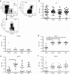
Recovery of LV-transduced human hematopoietic cells in the lymphoid organs of BRG-HIS mice. BRG-HIS mice generated with LV-transduced hHPC for the expression of shGag5 (grey diamonds, n = 5), shPol1 (black diamonds, n = 9), shPol47 (grey triangle, n = 17) or shR/T5 (black triangle, n = 11) were analyzed 10–13 weeks post-hHPC transplantation and compared with control animals (empty JS1-transduced hHPC; black circles, n = 26). (a) The flow cytometry dot plots show the transduction efficiency in vitro of hHPC transduced with LV (left panel, SSC side scatter) and the frequency of GFP expression among human hCD45+ hematopoietic cells in vivo in the BRG-HIS mice (right panel). (bf) The results are expressed as the GFP+ ratio between the frequency of human GFP+ cells measured in the animals (in vivo) and the frequency of GFP+ hHPC injected in the newborn mice (in vitro transduction efficiency). The GFP+ ratio is shown for the bone marrow (b), thymus (c), spleen (d), liver (e), and blood (f). The results (mean) are pooled from two to eight independent experiments, and each dot represents an individual animal. *P < 0.05; **P < 0.01 (Kruskall–Wallis test).
Figure 4
Figure 4
Differential shRNA expression in human cell subsets of BRG-HIS mice. GFP expression by several human immune cells was measured in the spleen of BRG-HIS mice. (a) The analyzed subsets were human T cells (CD3+), B cells (BDCA2CD19+), plasmacytoid dendritic cells (pDC; CD19BDCA2+), and monocytes (Mono; CD19BDCA2CD14+). (bg) GFP expression by these subsets was measured in BRG-HIS mice reconstituted with the following LV: control JS1 (b), shGag5 (c), shPol1 (d), shR/T5 (e), or shPol47 (f). The results are expressed as the GFP+ ratio between the frequency of human GFP+ cells measured in the spleen of the animals (in vivo) and the frequency of GFP+ hHPC injected in the newborn mice (in vitro transduction efficiency). The results (mean) are pooled from two to eight independent experiments, and each dot represents an individual animal. *P < 0.05; **P < 0.01 (Friedman test).
Figure 5
Figure 5
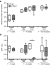
Impaired seeding of BRG-HIS mouse thymus by shPol47-expressing human progenitor cells. CD34+CD38 hHPC were transduced with JS1-control or shPol47-expressing LV and injected into newborn BRG mice. At 1 and 4 weeks post-hHPC transplantation, JS1 (white boxes; n = 4) and shPol47 (grey boxes; n = 5) BRG-HIS mice were analyzed in the major sites of hematopoiesis for the absolute number of human CD45+ cells (a) and for GFP+ ratio in human hematopoietic cells (b). The GFP+ ratio is the ratio between the frequency of human GFP+ cells measured in the animals (in vivo) and the frequency of GFP+ hHPC injected in the newborn mice (in vitro transduction efficiency). The results are pooled from four independent experiments, and each dot represents an individual animal. *P < 0.05 (Mann–Whitney test). BM, bone marrow; LIV, liver; THY, thymus.
Figure 6
Figure 6
In vitro and in vivo safety of combinatorial shRNA treatment. (a) Schematic representation of the R3 and R4 LV. The R3 vector expresses the three validated shRNAs (shR/T5, shPol1, and shPol47) and the R4 vector also contains shGag5. (b) Human fetal liver CD34+CD38 hHPC were transduced with JS1, R3 or R4-expressing LV and analyzed as in Figure 2b. (cg) JS1 (black circles, n = 10), R3 (grey squares, n = 7), R4 (black squares, n = 3) groups of BRG-HIS mice were analyzed 10–13 weeks after transplantation. The GFP+ ratio in human hematopoietic cells was determined in the bone marrow (c), thymus (d), spleen (e), liver (f), and blood (g). The GFP+ ratio is the ratio between the frequency of human GFP+ cells measured in the animals (in vivo) and the frequency of GFP+ hHPC injected in the newborn mice (in vitro transduction efficiency). (h) GFP expression by human immune cell subsets was measured in the spleen of BRG-HIS mice generated with R3-expressing hHPC. The results are pooled from two independent experiments, and each dot represents an individual animal. *P < 0.05; **P < 0.01 (Kruskall–Wallis test for group comparison and Friedman test for cell subset comparison).
Figure 7
Figure 7
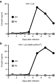
R3+ human CD4+ T cells from NSG-HIS mice inhibit HIV-1 replication ex vivo. Transduced (GFP+) human CD4+ T cells from spleen of JS1 and R3-treated NSG-HIS mice were sorted and infected either with HIV-1 LAI (a) or a HIV-1 LAI strain resistant to shPol1 (HIV-1 LAI D30N) (b). Virus spread was monitored by measuring CA-p24 production with ELISA. The HIV-1 inhibition experiment was repeated three-times and one representative experiment is shown.

References

    1. Yukl SA, Shergill AK, McQuaid K, Gianella S, Lampiris H, Hare CB, et al. Effect of raltegravir-containing intensification on HIV burden and T-cell activation in multiple gut sites of HIV-positive adults on suppressive antiretroviral therapy. AIDS. 2010;24:2451–2460. - PMC - PubMed
    1. Llibre JM, Buzón MJ, Massanella M, Esteve A, Dahl V, Puertas MC, et al. Treatment intensification with raltegravir in subjects with sustained HIV-1 viraemia suppression: a randomized 48-week study. Antivir Ther (Lond) 2012;17:355–364. - PubMed
    1. Siliciano JD, Kajdas J, Finzi D, Quinn TC, Chadwick K, Margolick JB, et al. Long-term follow-up studies confirm the stability of the latent reservoir for HIV-1 in resting CD4+ T cells. Nat Med. 2003;9:727–728. - PubMed
    1. Hütter G, Nowak D, Mossner M, Ganepola S, Müssig A, Allers K, et al. Long-term control of HIV by CCR5 Delta32/Delta32 stem-cell transplantation. N Engl J Med. 2009;360:692–698. - PubMed
    1. Allers K, Hütter G, Hofmann J, Loddenkemper C, Rieger K, Thiel E, et al. Evidence for the cure of HIV infection by CCR5?32/?32 stem cell transplantation. Blood. 2011;117:2791–2799. - PubMed