. 2014 Jan;12(1):143-154.
doi: 10.1158/1541-7786.MCR-13-0184. Epub 2013 Sep 3.

Integrin α3β1 can function to promote spontaneous metastasis and lung colonization of invasive breast carcinoma

Affiliations

Integrin α3β1 can function to promote spontaneous metastasis and lung colonization of invasive breast carcinoma

Bo Zhou et al. Mol Cancer Res. 2014 Jan.

Abstract

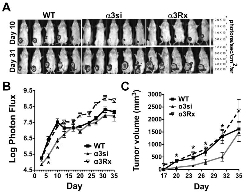
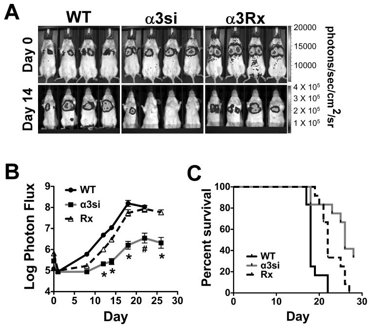
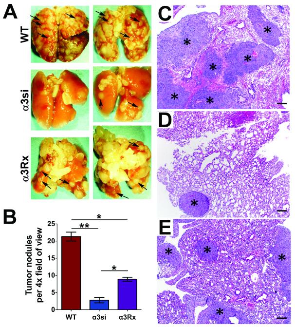
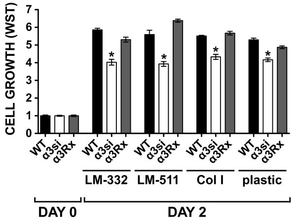
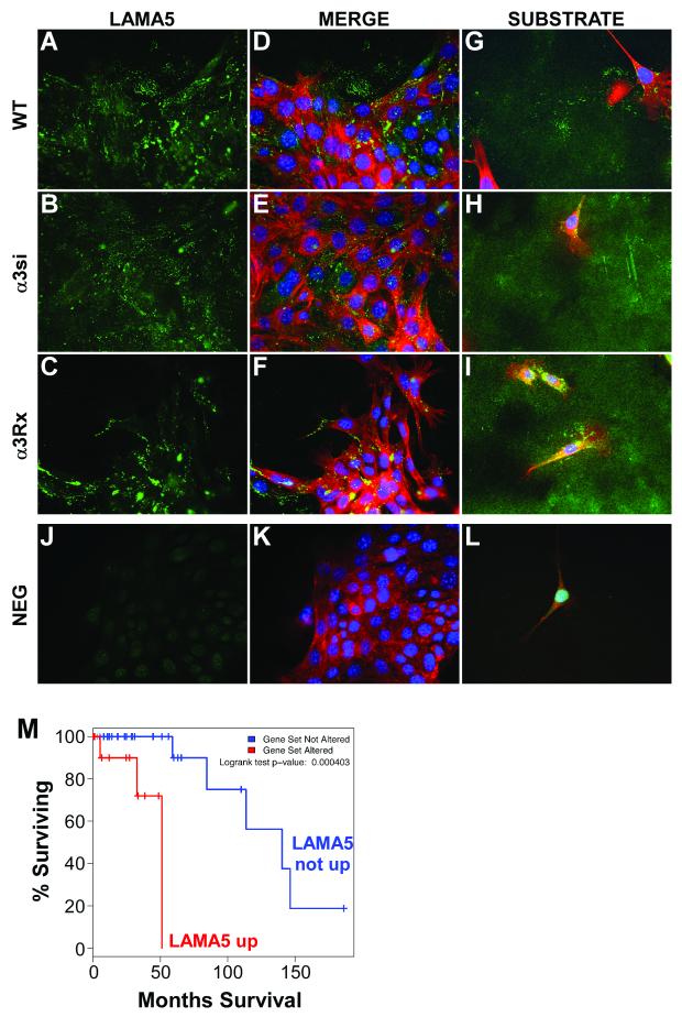
Significant evidence implicates α3β1 integrin in promoting breast cancer tumorigenesis and metastasis-associated cell behaviors in vitro and in vivo. However, the extent to which α3β1 is actually required for breast cancer metastasis remains to be determined. We used RNA interference to silence α3 integrin expression by approximately 70% in 4T1 murine mammary carcinoma cells, a model of aggressive, metastatic breast cancer. Loss of α3 integrin reduced adhesion, spreading, and proliferation on laminin isoforms, and modestly reduced the growth of orthotopically implanted cells. However, spontaneous metastasis to lung was strikingly curtailed. Experimental lung colonization after tail vein injection revealed a similar loss of metastatic capacity for the α3-silenced (α3si) cells, suggesting that critical, α3-dependent events at the metastatic site could account for much of α3β1's contribution to metastasis in this model. Reexpressing α3 in the α3si cells reversed the loss of metastatic capacity, and silencing another target, the small GTPase RhoC, had no effect, supporting the specificity of the effect of silencing α3. Parental, α3si, and α3-rescued cells, all secreted abundant laminin α5 (LAMA5), an α3β1 integrin ligand, suggesting that loss of α3 integrin might disrupt an autocrine loop that could function to sustain metastatic growth. Analysis of human breast cancer cases revealed reduced survival in cases where α3 integrin and LAMA5 are both overexpressed.

Implications: α3 integrin or downstream effectors may be potential therapeutic targets in disseminated breast cancers, especially when laminin α5 or other α3 integrin ligands are also over-expressed.


Figures

Figure 1
Figure 1. Impaired adhesion and spreading of α3 integrin-silenced 4T1 carcinoma cells
(A) Cell surface biotinylated wild type (WT), α3-silenced (α3si), and α3-rescued (α3Rx) 4T1 cells were lysed in 1% Triton X-100 detergent and α3β1 integrin was immunoprecipitated using the A3-CYT polyconal antibody (lanes 1-3) or the 9E10 anti-myc epitope antibody (lanes 4-6). The blot was visualized with DyLight 800-Neutravidin. Arrows indicate the α3 and β1 integrin bands. (B) Quantification of multiple independent blots by LI-COR infrared fluorescent scanner. Data are presented as % wild type expression ± SEM, n=6 blots. (C) Wild type, α3si, and α3Rx 4T1 cells were plated in wells with different coating concentrations of LM-332. After 25 min, non-adherent cells were removed by rinsing, and adherent cells were quantified by crystal violet staining. Compared to wild type or α3Rx cells, the α3si cells showed significantly reduced adhesion on wells coated 0.5 μg/ml and 1.0 μg/ml LM-332 (*P<0.001, ANOVA with Tukey post-test, n=4 wells/condition). (D) Wild type, α3si, and α3Rx 4T1 cells were plated on wells coated with 20 μg/ml collagen I or in BSA-blocked negative control wells. After 25 min, adherent cells were quantified as in (A). (E-G) Wild type, α3si, and α3Rx 4T1 cells were plated on glass coverslips coated with 1 μg/ml LM-332 for 30 min and then fixed and photographed with differential interference microscopy. (H) The spread area of wild type, α3si, and α3Rx cells was quantified with ImageJ software, as described in Materials and Methods. The spread area of α3si cells was significantly less than wild type or α3Rx cells (*P<0.01, ANOVA with Tukey post test, n≥98 cells per cell type).
Figure 2
Figure 2. Growth of primary tumors and total tumor burden
On day 0, 5,000 luciferase-expressing 4T1 wild type, α3si, and α3Rx cells were implanted in the 4th mammary fat pad of female Balb/C mice. (A) Bioluminescence imaging (BLI) of the cells on day 10 (when α3si tumor cell burden appeared reduced compared to controls) and day 31 (when α3si tumor cell burden appeared similar to wild type). (B) Total apparent tumor burden, measured as log photon flux, for the entire timecourse of the experiment. The α3si tumor cell burden was significantly less than wild type or α3Rx on days 6 and 10 (*P<0.001 vs wild type, P<0.01 vs α3Rx, ANOVA with Tukey post-test; n=10 mice per group). The slight reduction in photon flux observed on day 35 was due to the loss of some of the mice with the highest tumor burdens between day 31 and day 35. (C) Tumor volumes measured by caliper. The mean volume of the α3si tumors was significantly less than the volumes of both the wild type and α3Rx tumors from day 20 through day 31 (*P<0.01 on day 20, and P<0.05 on days 24-31, ANOVA with Tukey post-test, except for day 27, when α3si was significantly different from α3Rx, but not wild type).
Figure 3
Figure 3. Spontaneous metastasis is significantly impaired in α3 integrin-silenced 4T1 cells
(A) Lung colonization as quantified in ex-vivo BLI images (See Figure S4 for representative images). The α3si 4T1 cell colonization was significantly reduced compared to wild type or α3Rx cell colonization (*P<0.05 vs wild type, P<0.001 vs α3Rx, ANOVA with Tukey post-test; n = 10-14 mice per group; bars indicate mean ± SEM). (B) Wild type, α3si, and α3Rx 4T1 cells were cell surface-biotinylated, and α3β1 integrin was recovered by immunoprecipitation from Triton X-100 lysates, as in Figure 1. For each set, the lane marked “parental” corresponds to the original cell line implanted into mammary fat pad at the beginning of the assay, and the lanes numbered 1-3 correspond to sublines recovered from lung explants at the end of the assay. Quantification of band intensities by LI-COR blot imager is indicated under each lane (in arbitrary intensity units). (C) The relationship between primary tumor volume and lung colonization measured by BLI is graphed for each cell type. Values for R2 and P from Pearson correlation tests are shown for each graph. In no case was there a significant correlation between primary tumor volume and lung colonization.
Figure 4
Figure 4. Experimental lung metastasis is significantly impaired in α3 integrin-silenced 4T1 cells
On day 0, 50,000 wild type, α3si, or α3Rx luciferase-expressing 4T1 cells were injected by tail vein into female Balb/C mice. (A) BLI imaging on day 0, immediately after tail vein injection and on day 14, about halfway through the assay. (B) Total tumor burden (log photon flux) as measured by BLI. Metastatic colonization by the α3si cells was significantly reduced compared to wild type or α3Rx cells from day 12 onward (*P<0.001 vs wild type and α3Rx cells, on days 12-20; #P<0.05 vs wild type and P<0.001 vs α3Rx on day 22; and *P<0.001 vs α3Rx on day 26, ANOVA with Tukey post-test, n=15 mice per group). (C) Survival to endpoint for mice bearing wild type, α3si, or α3Rx 4T1 cells. Survival of mice with α3si 4T1 cells was significantly increased compared to mice with wild type or α3Rx 4T1 cells (P<0.0001 vs wild type and P = 0.0127 vs α3Rx, Mantel-Cox log-rank test).
Figure 5
Figure 5. Gross and histologic analysis of experimental lung metastasis
(A) Lungs recovered 18-28 days after injection from mice bearing wild type, α3si, or a3Rx 4T1 cells were paraformaldehyde-fixed and photographed using a dissecting microscope prior to paraffin-embedding. Pulmonary metastases appear as pale tan nodules on the surfaces of lungs (arrows point to examples). Fewer nodules were evident on lungs from mice with α3si cells. (B) Quantification of tumor nodules per 4X field in H&E-stained sections from paraffin-embedded lungs. The α3si 4T1 cells formed fewer nodules than either wild type or α3Rx 4T1 cells. The wild type cells formed more nodules than the α3Rx cells, indicating a partial restoration of α3 function for the α3Rx cells in this assay. (*P<0.01, ANOVA with Tukey post-test, n=3 mice per group, 3 slides per mouse, 3 fields per slide, for a total of 27 fields per cell type). Shown at right are representative photomicrographs of lungs from mice injected with (C) wild type, (D) α3si, or (E) α3Rx 4T1 cells. Asterisks indicate examples of tumor nodules within the pulmonary parenchyma. HE, bars = 200 μm.
Figure 6
Figure 6. Impaired proliferation of α3-silenced 4T1 cells
Wild type, α3-silenced, and α3Rx 4T1 cells were plated in 2% FBS in wells coated with 1 μg/ml LM-332, 1 μg/ml LM-511, 10 μg/ml collagen I, or in uncoated wells. One set of wells was assayed by WST-1 on day 0 (the day of plating) to measure cells input, and the remaining wells were assayed on day 2. Bars represent mean ± SEM for 6 wells per cell type per condition, normalized to day 0 values. The growth of the α3si cells was significantly less than that of either the wild type or α3Rx cells on all substrates tested (*P<0.001, ANOVA with Tukey post-test).
Figure 7
Figure 7. Laminin α5 expression by 4T1 cells
(A-C) Wild type, α3si, and α3Rx 4T1 cells were cultured on collagen I-coated coverslips overnight, then fixed and stained with polyclonal anti-laminin α5 antibody (LAMA5). (D-F) The merged images show counterstaining for F-actin and nuclei with phalloidin and DAPI. (G-I) Longer exposure images in cell-free areas highlight laminin α5 deposition on the substrate. (J-L) Negative control staining with normal rabbit IgG. (M) Survival analysis of α3 integrin-overexpressing human breast cancer cases, based on whether or not laminin-α5 (LAMA5) is also upregulated. LAMA5 overexpressing cases showed reduced survival (P=0.000403, logrank test).

Similar articles

Cited by

References

    1. Weaver VM, Lelièvre S, Lakins JN, Chrenek MA, Jones JCR, Giancotti F, et al. beta4 integrin-dependent formation of polarized three-dimensional architecture confers resistance to apoptosis in normal and malignant mammary epithelium. Cancer Cell. 2002;2:205–16. - PMC - PubMed
    1. Zahir N, Lakins JN, Russell A, Ming W, Chatterjee C, Rozenberg GI, et al. Autocrine laminin-5 ligates alpha6beta4 integrin and activates RAC and NFkappaB to mediate anchorage-independent survival of mammary tumors. J Cell Biol. 2003;163:1397–407. - PMC - PubMed
    1. Plopper GE, Domanico SZ, Cirulli V, Kiosses WB, Quaranta V. Migration of breast epithelial cells on Laminin-5: differential role of integrins in normal and transformed cell types. Breast Cancer Res Treat. 1998;51:57–69. - PubMed
    1. Martin KJ, Kwan CP, Nagasaki K, Zhang X, O’Hare MJ, Kaelin CM, et al. Down-regulation of laminin-5 in breast carcinoma cells. Mol Med. 1998;4:602–13. - PMC - PubMed
    1. Henning K, Berndt A, Katenkamp D, Kosmehl H. Loss of laminin-5 in the epithelium-stroma interface: an immunohistochemical marker of malignancy in epithelial lesions of the breast. Histopathology. 1999;34:305–9. - PubMed

Publication types

MeSH terms