. 2013 Dec;41(22):10462-75.
doi: 10.1093/nar/gkt798. Epub 2013 Sep 3.

Single transcriptional and translational preQ1 riboswitches adopt similar pre-folded ensembles that follow distinct folding pathways into the same ligand-bound structure

Affiliations

Single transcriptional and translational preQ1 riboswitches adopt similar pre-folded ensembles that follow distinct folding pathways into the same ligand-bound structure

Krishna C Suddala et al. Nucleic Acids Res. 2013 Dec.

Abstract

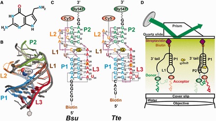
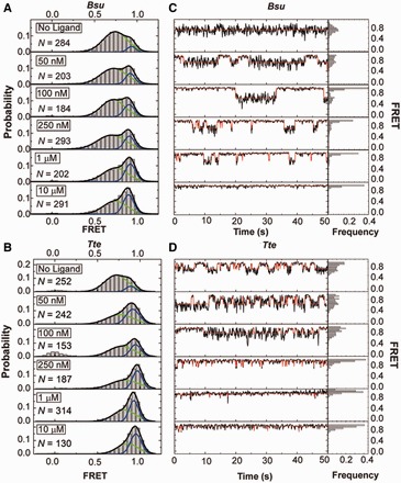
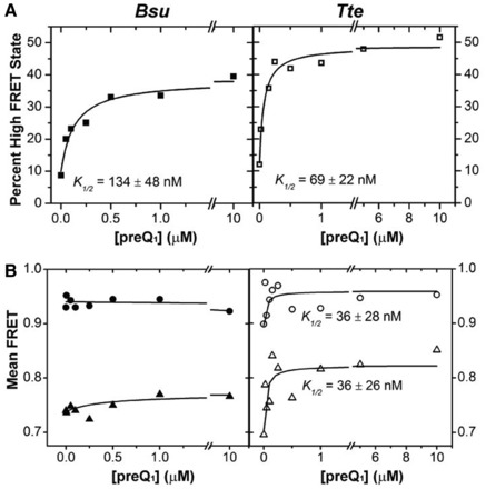
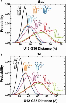
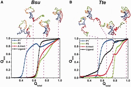
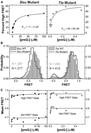
Riboswitches are structural elements in the 5' untranslated regions of many bacterial messenger RNAs that regulate gene expression in response to changing metabolite concentrations by inhibition of either transcription or translation initiation. The preQ1 (7-aminomethyl-7-deazaguanine) riboswitch family comprises some of the smallest metabolite sensing RNAs found in nature. Once ligand-bound, the transcriptional Bacillus subtilis and translational Thermoanaerobacter tengcongensis preQ1 riboswitch aptamers are structurally similar RNA pseudoknots; yet, prior structural studies have characterized their ligand-free conformations as largely unfolded and folded, respectively. In contrast, through single molecule observation, we now show that, at near-physiological Mg(2+) concentration and pH, both ligand-free aptamers adopt similar pre-folded state ensembles that differ in their ligand-mediated folding. Structure-based Gō-model simulations of the two aptamers suggest that the ligand binds late (Bacillus subtilis) and early (Thermoanaerobacter tengcongensis) relative to pseudoknot folding, leading to the proposal that the principal distinction between the two riboswitches lies in their relative tendencies to fold via mechanisms of conformational selection and induced fit, respectively. These mechanistic insights are put to the test by rationally designing a single nucleotide swap distal from the ligand binding pocket that we find to predictably control the aptamers' pre-folded states and their ligand binding affinities.


Figures

Figure 1.
Figure 1.
Structural comparison of the Bsu and Tte preQ1 riboswitches. (A) Structure of preQ1 (7-aminomethyl-7-deazaguanine). (B) Structural overlay of the Bsu (colored, PDB ID 3FU2, chain A) and Tte (gray, PDB ID 3Q50) riboswitch crystal structures. The sugar-phosphate backbone is shown as a single ribbon. preQ1 is space-filled and colored as in A. Secondary structure elements are color-coded as indicated. (C) Secondary structure maps of the Bsu and Tte riboswitches with interactions shown in Leontis–Westhof nomenclature (14). Individual secondary structures are color-coded as in (B), and the locations of fluorophores and biotin are indicated. (D) Prism-based TIRFM setup for smFRET.
Figure 2.
Figure 2.
smFRET characterization of single preQ1 riboswitch molecules. (A) smFRET histograms of the Bsu riboswitch with increasing ligand concentration as indicated; N, number of molecules sampled. Green and blue lines indicate Gaussian fits of the mid- and high-FRET states, respectively. Black lines indicate cumulative fits. (B) Same as in A, but for the Tte riboswitch. (C) Exemplary FRET time traces of the Bsu riboswitch for each condition. Idealized HMM fits are shown as red line. The population of each FRET state is shown as a frequency bar graph to the right. (D) Same as in C, but for the Tte riboswitch.
Figure 3.
Figure 3.
Effect of ligand on the distribution of the mid- and high-FRET states. (A) The FRET histograms of Figure 2 were quantified, and the percentage high-FRET state was plotted as a function of ligand concentration. The data were fit with a non-cooperative binding isotherm and the respective apparent K1/2 values are indicated for both Bsu (closed symbols) and Tte (open symbols). (B) The centers of the Gaussian fits for the mid-FRET (green) and high-FRET (blue) states from Figure 2 were plotted as a function of ligand concentration and fit with a non-cooperative binding isotherm, yielding the K1/2 values indicated for the Tte riboswitch.
Figure 4.
Figure 4.
Coarse grained TOPRNA simulations predicting distance distributions between the fluorophore labeled residues as a function of specific interactions in the Bsu (A) and Tte (B) riboswitches. Color code is as follows: green, stacked 3′ tail; red, unstacked 3′ tail; purple, blue, orange and cyan (A, only), partially docked into the P1 and/or P2 stem with varying degrees of intersegmental and stacking interactions as indicated; black, fully folded as found in the ligand-bound crystal structures (see Supplementary Tables S1, S2 and S4 for details).
Figure 5.
Figure 5.
Gō model simulations of single Bsu (A) and Tte (B) riboswitch molecules. Fraction of native contacts for each structural component, Qsec (P1, blue; P2, green; A-tract, red; ligand, black), averaged over each 51 simulations and plotted as a function of the fraction of total contacts observed in the native folded structure, Qtotal. Above, characteristic points along the folding pathway are illustrated with each one representative conformation.
Figure 6.
Figure 6.
smFRET characterization of riboswitch mutants. (A) The Gaussian distributions from Supplementary Figure S10 were quantified and the fraction high-FRET state was plotted as a function of preQ1 concentration for the Bsu (closed symbols) and Tte (open symbols) riboswitches. (B) smFRET histograms of the Bsu and Tte riboswitches in wild-type (WT, gray bars) and mutant (black line) forms, in the absence of preQ1. (C) The centers of the mid-FRET (triangles) and high-FRET (circles) states in Supplementary Figure S10 were plotted as a function of ligand concentration for both the Bsu (closed symbols) and Tte (open symbols) riboswitches.
Figure 7.
Figure 7.
Parsimonious folding model of the Bsu and Tte preQ1 riboswitches. A combination of smFRET and computational simulations support a model in which the preQ1 ligand binds late and concomitantly with the docking of the 3′ tail and formation of the P2 stem in the Bsu riboswitch, signifying conformational selection (represented in green). By contrast, early binding of the preQ1 ligand to a partially unfolded conformation induces folding into the bound structure of the Tte riboswitch, consistent with an induced fit model (represented in blue). Both mechanisms are not mutually exclusive, and it is plausible that a combination of both induced fit and conformational selection mechanisms are at work in both riboswitches (59). The size of the white circle and the gray outlines describe the extent of conformational heterogeneity of each state.

References

    1. Winkler WC, Nahvi A, Roth A, Collins JA, Breaker RR. Control of gene expression by a natural metabolite-responsive ribozyme. Nature. 2004;428:281–286. - PubMed
    1. Nudler E, Mironov AS. The riboswitch control of bacterial metabolism. Trends Biochem. Sci. 2004;29:11–17. - PubMed
    1. Mandal M, Breaker RR. Gene regulation by riboswitches. Nat. Rev. Mol. Cell Biol. 2004;5:451–463. - PubMed
    1. Winkler WC, Breaker RR. Regulation of bacterial gene expression by riboswitches. Annu. Rev. Microbiol. 2005;59:487–517. - PubMed
    1. Breaker RR. Prospects for riboswitch discovery and analysis. Mol. Cell. 2011;43:867–879. - PMC - PubMed

Publication types