

Wounded embryonic corneas exhibit nonfibrotic regeneration and complete innervation
- PMID: 24003085
- PMCID: PMC3783042
- DOI: 10.1167/iovs.13-12504
Wounded embryonic corneas exhibit nonfibrotic regeneration and complete innervation
Abstract
Purpose: Wound healing in adult corneas is characterized by activation of keratocytes and extracellular matrix (ECM) synthesis that results in fibrotic scar formation and loss of transparency. Since most fetal wounds heal without scaring, we investigated the regenerative potential of wounded embryonic corneas.
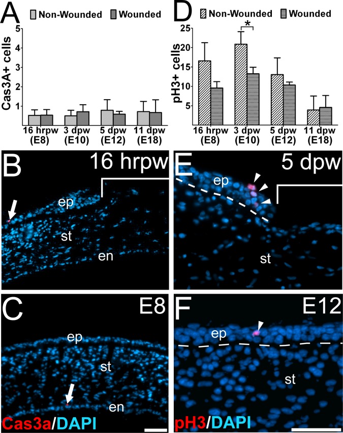
Methods: On embryonic day (E) 7 chick corneas were wounded by making a linear incision traversing the epithelium and anterior stroma. Wounded corneas were collected between E7 and E18, and analyzed for apoptosis, cell proliferation, staining of ECM components, and corneal innervation.
Results: Substantial wound retraction was observed within 16-hours postwounding (hpw) and partial re-epithelialized by 5-days postwounding (dpw). Corneal wounds were fully re-epithelialized by 11 dpw with no visible scars. There was no difference in the number of cells undergoing apoptosis between wounded and control corneas. Cell proliferation was reduced in the wounded corneas, albeit mitotic cells in the regenerating epithelium. Staining for alpha-smooth muscle actin (α-SMA), tenascin, and fibronectin was vivid but transient at the wound site. Staining for procollagen I, perlecan, and keratan sulfate proteoglycan was reduced at the wound site. Wounded corneas were fully regenerated by 11 dpw and showed similar patterns of staining for ECM components, albeit an increase in perlecan staining. Corneal innervation was inhibited during wound healing, but regenerated corneas were innervated similar to controls.
Conclusions: These data show that minimal keratocyte activation, rapid ECM reconstruction, and proper innervation occur during nonfibrotic regeneration of the embryonic cornea.
Keywords: corneal development; corneal wound healing; extracellular matrix; innervation.
Figures


Similar articles
-
Recapitulation of normal collagen architecture in embryonic wounded corneas.Sci Rep. 2020 Aug 14;10(1):13815. doi: 10.1038/s41598-020-70658-y. Sci Rep. 2020. PMID: 32796881 Free PMC article.
-
Investigating Scarless Tissue Regeneration in Embryonic Wounded Chick Corneas.J Vis Exp. 2022 May 2;(183). doi: 10.3791/63570. J Vis Exp. 2022. PMID: 35575534
-
Altered KSPG expression by keratocytes following corneal injury.Mol Vis. 2003 Nov 21;9:615-23. Mol Vis. 2003. PMID: 14654769
-
The corneal fibrosis response to epithelial-stromal injury.Exp Eye Res. 2016 Jan;142:110-8. doi: 10.1016/j.exer.2014.09.012. Exp Eye Res. 2016. PMID: 26675407 Free PMC article. Review.
-
Role of aquaporins in corneal healing post chemical injury.Exp Eye Res. 2023 Mar;228:109390. doi: 10.1016/j.exer.2023.109390. Epub 2023 Jan 22. Exp Eye Res. 2023. PMID: 36696947 Free PMC article. Review.
Cited by
-
Recapitulation of normal collagen architecture in embryonic wounded corneas.Sci Rep. 2020 Aug 14;10(1):13815. doi: 10.1038/s41598-020-70658-y. Sci Rep. 2020. PMID: 32796881 Free PMC article.
-
Sema3A maintains corneal avascularity during development by inhibiting Vegf induced angioblast migration.Dev Biol. 2014 Jul 15;391(2):241-50. doi: 10.1016/j.ydbio.2014.04.017. Epub 2014 May 6. Dev Biol. 2014. PMID: 24809797 Free PMC article.
-
Semi-autonomous wound invasion via matrix-deposited, haptotactic cues.J Theor Biol. 2023 Jul 7;568:111506. doi: 10.1016/j.jtbi.2023.111506. Epub 2023 Apr 22. J Theor Biol. 2023. PMID: 37094713 Free PMC article.
-
Matricellular proteins in the trabecular meshwork: review and update.J Ocul Pharmacol Ther. 2014 Aug;30(6):447-63. doi: 10.1089/jop.2014.0013. Epub 2014 Jun 5. J Ocul Pharmacol Ther. 2014. PMID: 24901502 Free PMC article. Review.
-
Mesenchymal proteases and tissue fluidity remodel the extracellular matrix during airway epithelial branching in the embryonic avian lung.Development. 2019 Aug 19;146(16):dev175257. doi: 10.1242/dev.175257. Development. 2019. PMID: 31371376 Free PMC article.
References
-
- Ghiasi H, Cai S, Perng GC, Nesburn AB, Wechsler SL. The role of natural killer cells in protection of mice against death and corneal scarring following ocular HSV-1 infection. Antiviral Res. 2000; 45: 33–45 - PubMed
-
- Hassell JR, Cintron C, Kublin C, Newsome DA. Proteoglycan changes during restoration of transparency in corneal scars. Arch Biochem Biophys. 1983; 222: 362–369 - PubMed
-
- Wilson SE, Mohan RR, Mohan RR, Ambrósio R Jr, Hong JW, Lee JS. The corneal wound healing response: cytokine-mediated interaction of the epithelium, stroma, and inflammatory cells. Prog Retin Eye Res. 2001; 20: 625–637 - PubMed
-
- Jester JV, Petroll WM, Barry PA, Cavanagh HD. Expression of alpha-smooth muscle (alpha-SM) actin during corneal stromal wound healing. Invest Ophthalmol Vis Sci. 1995; 36: 809–819 - PubMed
-
- Matsubara M, Girard MT, Kublin CL, Clinton C, Fini ME. Differential roles for two gelatinolytic enzymes of the matrix metalloproteinase family in the remodeling cornea. Dev Biol. 1991; 147: 425–439 - PubMed
Publication types
MeSH terms
Grants and funding
LinkOut - more resources
Full Text Sources
Other Literature Sources
Medical
