. 2013 Sep 17;110(38):15360-4.
doi: 10.1073/pnas.1302218110. Epub 2013 Sep 3.

How minute sooglossid frogs hear without a middle ear

Affiliations

How minute sooglossid frogs hear without a middle ear

Renaud Boistel et al. Proc Natl Acad Sci U S A. .

Abstract

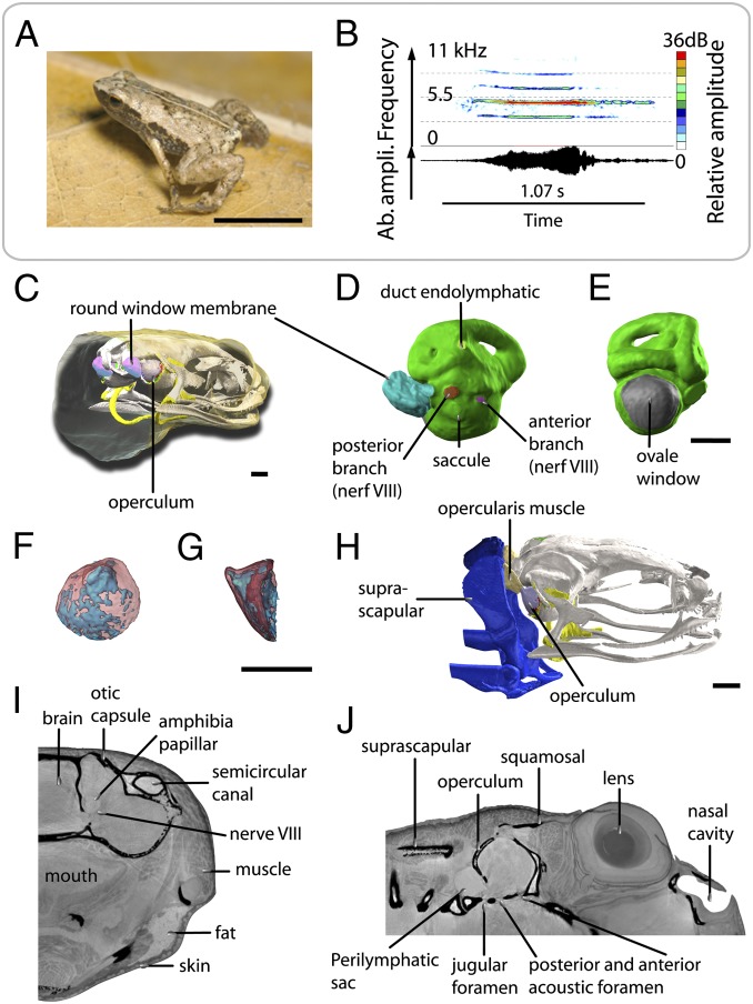
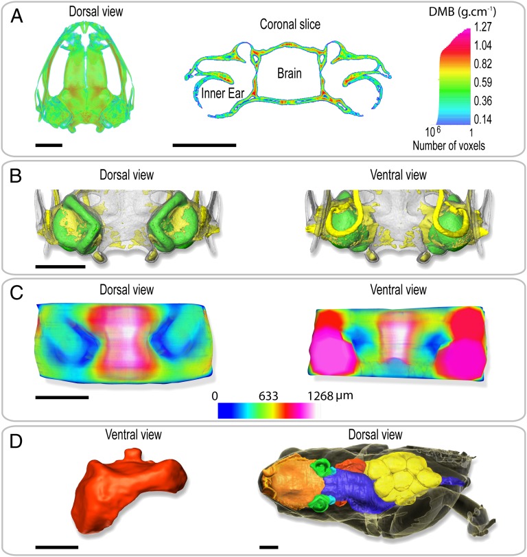
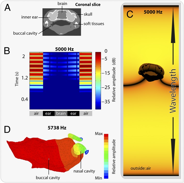
Acoustic communication is widespread in animals. According to the sensory drive hypothesis [Endler JA (1993) Philos Trans R Soc Lond B Biol Sci 340(1292):215-225], communication signals and perceptual systems have coevolved. A clear illustration of this is the evolution of the tetrapod middle ear, adapted to life on land. Here we report the discovery of a bone conduction-mediated stimulation of the ear by wave propagation in Sechellophryne gardineri, one of the world's smallest terrestrial tetrapods, which lacks a middle ear yet produces acoustic signals. Based on X-ray synchrotron holotomography, we measured the biomechanical properties of the otic tissues and modeled the acoustic propagation. Our models show how bone conduction enhanced by the resonating role of the mouth allows these seemingly deaf frogs to communicate effectively without a middle ear.

Keywords: X-ray imaging; audition; earless frog; extra-tympanic pathways.


Conflict of interest statement

The authors declare no conflict of interest.

Figures

Fig. 1.
Fig. 1.
Acoustic behavior and volume rendering of ear region of S. gardineri. (A) Picture of one adult male of S. gardineri. (B) Oscillogram (Lower) and sonogram (Upper) of the advertisement call. (C) 3D visualization of the operculum and endolymphatic sac of the oval window in the head, in 3/4 posterior view with the skin made transparent. (D) Median view of the inner ear (in green) with the endolymphatic sac connected to the left round window, showing the location of the posterior (red) and anterior (violet) branches of nerve VIII, the endolymphatic duct (pale green), and extracranial lymphatic sac (blue-green). (E) Posterior view of the left inner ear, showing the surface occupied by the oval window (in gray). (F and G) 3D visualization of the operculum in transparency (bone in red, cartilage in blue): posterior (F) and lateral (G) views. (H) 3D 3/4 lateral view of the opercular system with the opercular muscle shown in pink. (I and J) Sagittal and frontal sections of holotomographic images showing the foramen of the otic capsule, the innervation of the inner ear, and the organization of the tissues surrounding the ear. (Scale bar, 0.5 mm.)
Fig. 2.
Fig. 2.
Volume rendering of holotomography of the body and the tissues surrounding the ear of S. gardineri. (A) 3D map (Left) and virtual sagittal section (Right) illustrating the degree of mineralization of the skull as determined by absorption tomography. (B) Visualization of the degree of ossification of the otic region of the skull in dorsal (Left) and ventral (Right) views. The skull is transparent, cartilage is shown in yellow, and the inner ears in green. (C) 3D map of the thickness of the tissues separating the inner ears and the gaseous medium. (Left) Dorsal view and (Right) ventral view of the oral region. The thinnest parts are shown in dark blue. (D) 3D visualization of the pulmonary system of a gravid female (SVL = 11.65 mm). Red, pulmonary and laryngeal cavity in ventral view. To right, dorsal view of female in transparency visualizing the anatomy and the volume occupied by the nasal and oral cavities (orange), the inner ears (green), the digestive system (blue), the ovarian masses (yellow), and the pulmonary system. Note the left forelimb was removed for molecular analysis. (Scale bar, 1 mm.)
Fig. 3.
Fig. 3.
Bone conduction in S. gardineri. (A) Image segmentation at the level of the inner ears used for bone conduction simulations. (B) Graphic representation of the acoustic pressure as a function of time (ms) along a line crossing the two inner ears; color bar shows the relative pressure amplitude in decibels. (C) Snapshot of the simulated field: visualization of the propagation of a pressure wave (at 5 kHz) on a coronal section of the skull. (D) Finite-element acoustic mode of the oral cavity and the nasal cavities with open nostrils in a 3/4 dorsal view. Color scale represents the acoustic pressure in Pascals.

Similar articles

Cited by

References

    1. Schnupp JWH, Carr CE. On hearing with more than one ear: Lessons from evolution. Nat Neurosci. 2009;12(6):692–697. - PMC - PubMed
    1. Allen J. Cochlear modeling. IEEE ASSP Mag. 1985;2(1):3–20.
    1. Clack JA. Discovery of the earliest-known tetrapod stapes. Nature. 1989;342(6248):425–427. - PubMed
    1. Van Bergeijk WA. Evolution of the sense of hearing in vertebrates. Am Zool. 1966;6(3):371–377. - PubMed
    1. Jaslow AP, Hetherington TE, Lombard RE. In: The Evolution of the Amphibian Auditory System. Fritzsch B, Ryan MJ, Wilczynski W, Hetherington TE, Walkowiak W, editors. New York: Wiley; 1988. pp. 69–91.