Review
. 2013 Sep 1;5(9):a008706.
doi: 10.1101/cshperspect.a008706.

Mitochondrial regulation of cell death

Affiliations
Review

Mitochondrial regulation of cell death

Stephen W G Tait et al. Cold Spring Harb Perspect Biol. .

Abstract

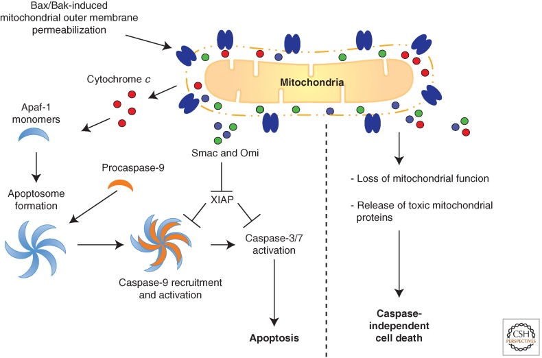
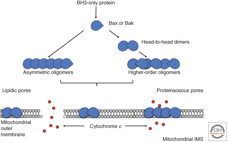
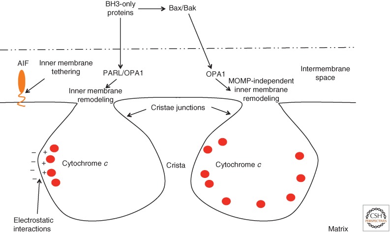
Although required for life, paradoxically, mitochondria are often essential for initiating apoptotic cell death. Mitochondria regulate caspase activation and cell death through an event termed mitochondrial outer membrane permeabilization (MOMP); this leads to the release of various mitochondrial intermembrane space proteins that activate caspases, resulting in apoptosis. MOMP is often considered a point of no return because it typically leads to cell death, even in the absence of caspase activity. Because of this pivotal role in deciding cell fate, deregulation of MOMP impacts on many diseases and represents a fruitful site for therapeutic intervention. Here we discuss the mechanisms underlying mitochondrial permeabilization and how this key event leads to cell death through caspase-dependent and -independent means. We then proceed to explore how the release of mitochondrial proteins may be regulated following MOMP. Finally, we discuss mechanisms that enable cells sometimes to survive MOMP, allowing them, in essence, to return from the point of no return.


Figures

Figure 1.
Figure 1.
Mitochondrial regulation of cell death. Bax/Bak-mediated mitochondrial outer membrane permeabilization (MOMP) can lead to caspase-dependent apoptosis (left) or caspase-independent cell death (right). Following MOMP, soluble proteins are released from the mitochondrial intermembrane space into the cytoplasm. Cytochrome c binds to monomeric Apaf-1 leading to its conformational change and oligomerization. Procaspase-9 is recruited to heptameric Apaf-1 complexes forming the apoptosome. This leads to activation of caspase-9 and, through caspase-9-mediated cleavage, activation of the executioner caspases-3 and -7. Release of Smac and Omi from the mitochondrial intermembrane space facilitates caspase activation by neutralizing the caspase inhibitor XIAP. MOMP can also lead to nonapoptotic cell death through a gradual loss of mitochondrial function and/or release of mitochondrial proteins that kill the cell in a caspase-independent manner.
Figure 2.
Figure 2.
Mechanism of Bax/Bak activation and MOMP. BH3 domain-only proteins directly bind and activate Bax and Bak. Activated Bax and Bak form higher-order oligomers, either through asymmetric oligomers (Bax) or through the formation of higher-order oligomers formed by head-to-head Bax or Bak dimers. How oligomeric Bax and Bak permeabilize the mitochondrial outer membrane is unclear. Two prominent models argue that Bax and Bak do this either by inducing lipidic pores (left) or by directly forming proteinaceous pores (right).
Figure 3.
Figure 3.
Post-MOMP regulation of mitochondrial intermembrane space protein release. The intermembrane space protein AIF is tethered to the mitochondrial inner membrane and requires cleavage to liberate it from the mitochondria upon MOMP. The majority of cytochrome c is sequestered within mitochondrial cristae; electrostatic interactions facilitate its association with the inner membrane. Some studies argue that cristae remodeling must occur to allow cytochrome c egress from the mitochondrial cristae following MOMP. Cristae remodeling can occur in a MOMP-independent manner by BH3 proteins (in a Bax/Bak-independent manner) or by activated Bax and Bak. Remodeling is dependent upon the intermembrane space rhomboid protease PARL and the dynamin-like GTPase OPA1.
Figure 4.
Figure 4.
Regulation of apoptosome activity. Various molecules, including tRNA, potassium, and ATP, can competitively inhibit cytochrome c–Apaf-1 interactions, thereby blocking apoptosome formation. Apaf-1 oligomerization can be positively affected by proteins such as PHAP that facilitate nucleotide exchange, whereas intracellular calcium levels inhibit this event. Various proteins, including heat shock proteins (Hsps) and kinases such as Rsk can directly inhibit Apaf-1 oligomerization through interaction with Apaf-1 or by inhibitory phosphorylation. The activity of the apoptosome can also be inhibited by the kinase activity of Erk1/2 and Cdk-1. Finally, proteins such as PCID1 can regulate the intracellular levels of procaspase-9, thereby regulating apoptosome activity.

References

    1. Abdelwahid E, Yokokura T, Krieser RJ, Balasundaram S, Fowle WH, White K 2007. Mitochondrial disruption in Drosophila apoptosis. Dev Cell 12: 793–806 - PubMed
    1. Albeck JG, Burke JM, Aldridge BB, Zhang M, Lauffenburger DA, Sorger PK 2008. Quantitative analysis of pathways controlling extrinsic apoptosis in single cells. Mol Cell 30: 11–25 - PMC - PubMed
    1. Allan LA, Clarke PR 2007. Phosphorylation of caspase-9 by CDK1/cyclin B1 protects mitotic cells against apoptosis. Mol Cell 26: 301–310 - PubMed
    1. Allan LA, Morrice N, Brady S, Magee G, Pathak S, Clarke PR 2003. Inhibition of caspase-9 through phosphorylation at Thr 125 by ERK MAPK. Nat Cell Biol 5: 647–654 - PubMed
    1. Antonsson B, Conti F, Ciavatta A, Montessuit S, Lewis S, Martinou I, Bernasconi L, Bernard A, Mermod JJ, Mazzei G, et al. 1997. Inhibition of Bax channel-forming activity by Bcl-2. Science 277: 370–372 - PubMed

MeSH terms

LinkOut - more resources