Review
. 2013 Sep 1;5(9):a012716.
doi: 10.1101/cshperspect.a012716.

DNA damage sensing by the ATM and ATR kinases

Affiliations
Review

DNA damage sensing by the ATM and ATR kinases

Alexandre Maréchal et al. Cold Spring Harb Perspect Biol. .

Abstract

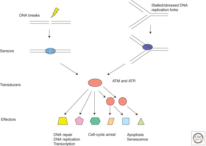
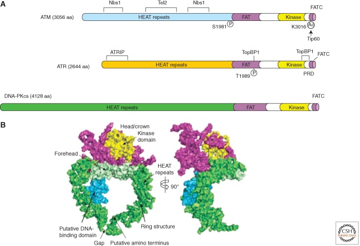
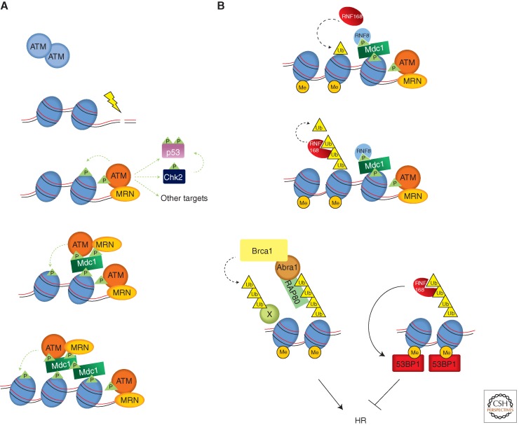
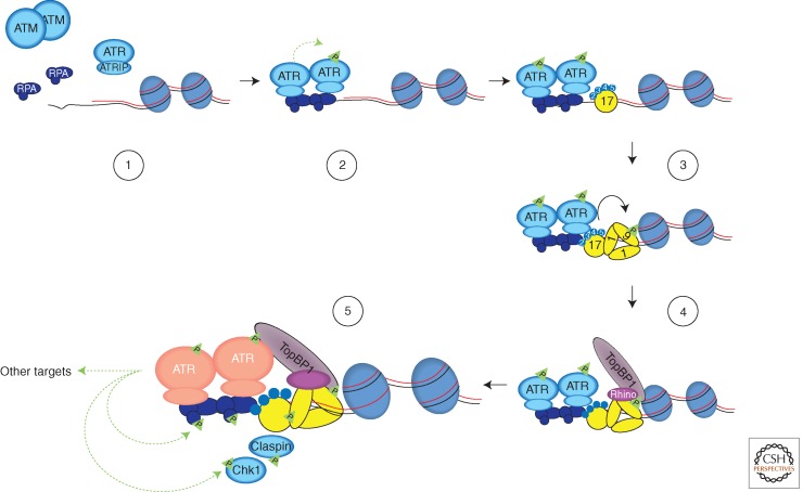
In eukaryotic cells, maintenance of genomic stability relies on the coordinated action of a network of cellular processes, including DNA replication, DNA repair, cell-cycle progression, and others. The DNA damage response (DDR) signaling pathway orchestrated by the ATM and ATR kinases is the central regulator of this network in response to DNA damage. Both ATM and ATR are activated by DNA damage and DNA replication stress, but their DNA-damage specificities are distinct and their functions are not redundant. Furthermore, ATM and ATR often work together to signal DNA damage and regulate downstream processes. Here, we will discuss the recent findings and current models of how ATM and ATR sense DNA damage, how they are activated by DNA damage, and how they function in concert to regulate the DDR.


Figures

Figure 1.
Figure 1.
The framework of the DDR signaling pathway. Like other signal transduction pathways, the DDR signaling pathway consists of signal sensors, transducers, and effectors. The sensors of this pathway are proteins that recognize DNA structures induced by DNA damage and DNA replication stress. The transducers of this pathway are kinases, including ATM, ATR, and their downstream kinases. The effectors of this pathway are substrates of ATM, ATR, and their downstream kinases. These effectors of ATM and ATR are involved in a broad spectrum of cellular processes that are important for maintenance of genomic stability of organisms.
Figure 2.
Figure 2.
Structural outlines of PIKKs. (A) Schematic of the functional domains and selected posttranslational modifications of ATM, ATR, and DNA-PKcs. The bulk of ATM and ATR are composed of a large number (40–50) of α-helical HEAT repeats. Some of these repeats are involved in the interaction with other proteins. The kinase domains of ATM and ATR are located near their carboxyl termini, and are flanked by FAT and FATC domains. ATM and ATR are autophosphorylated in or near their FAT domains after DNA damage, which regulates their activation. The acetylation of the carboxyl terminus of ATM by Tip60 is also involved in ATM activation. A PIKK regulatory domain (PRD) between the kinase and FATC domains of ATR was shown to modulate ATR activation. (B) Overall view of the DNA-PKcs structure. The front view (left) and side view (right) of DNA-PKcs. The various regions of DNA-PKcs are colored and labeled. (Panel B is from Sibanda et al. 2009; reprinted, with permission, from Nature Publishing Group © 2009.)
Figure 3.
Figure 3.
Activation of ATM by DSBs. (A) Recognition of DNA ends and chromatin by ATM. The MRN complex functions as a sensor of DNA ends and activates ATM. The ATM activated by DNA ends (red) phosphorylates substrates such as Chk2 and p53, and the H2AX in flanking nucleosomes. Phosphorylated H2AX (γH2AX) is recognized by Mdc1, which triggers a feed-forward loop that spreads activated ATM and γH2AX over large chromatin domains. (B) A ubiquitination cascade at DSBs that promotes the recruitment of Brca1 and 53BP1. The E3 ubiquitin ligase RNF8 is recruited to DSBs via phosphorylated Mdc1. RNF8 promotes another E3 ligase RNF168 to ubiquitinate substrates including histones H2A and H2AX. With the help of the HECT domain ligase HERC2 (not depicted here), RNF168 extends the K63-linked ubiquitin chains on substrates. These ubiquitin chains are recognized by RAP80, which forms a complex with Brca1 through Abraxas. Brca1, which is also a ubiquitin ligase, is critical for HR. RNF168 also promotes the recruitment of 53BP1, which binds the dimethylated lysine 20 of histone H4 (H4K20me2) exposed around DSBs. In contrast to Brca1, 53BP1 antagonizes HR. How the recruitment of Brca1 and 53BP1 is coordinated remains unknown. The dashed green and black lines represent phosphorylation and ubiquitylation events, respectively.
Figure 4.
Figure 4.
A fail-safe, multistep mechanism for ATR activation. Increased amounts of ssDNA are generated by resection of DNA ends or by uncoordinated DNA unwinding and synthesis at replication forks. Extensively resected DNA ends are no longer recognized by ATM efficiently. Once coated by RPA, ssDNA recruits the ATR-ATRIP complex (1), and promotes ATR trans-autophosphorylation (2). RPA-ssDNA also promotes the recruitment of the Rad17-Rfc2-5 clamp loader to junctions between ssDNA and dsDNA, and the loading of Rad9-Rad1-Hus1 (9-1-1) checkpoint clamps onto dsDNA (3). TopBP1 interacts with phosphorylated Rad9 and with Rhino, which associates with 9-1-1 (4). The TopBP1 recruited to dsDNA by 9-1-1 and Rhino engages the ATR-ATRIP complex on RPA-ssDNA through the ATR autophosphorylation site T1989. This process enables TopBP1 to stimulate ATR-ATRIP to its full capacity (pink) on ssDNA (5). TopBP1 may also function as a scaffold to facilitate ATR substrate recognition. This multistep process for ATR activation ensures that ATR is only activated when both ssDNA and ssDNA/dsDNA junctions are present at sites of DNA damage and are recognized by DNA damage sensors, providing a fail-safe but versatile mechanism to signal DNA damage. The dashed green lines represent phosphorylation events, and the solid black line represents the loading of 9-1-1 by the Rad17-RFC2-5 complex.

References

    1. Adams KE, Medhurst AL, Dart DA, Lakin ND 2006. Recruitment of ATR to sites of ionising radiation-induced DNA damage requires ATM and components of the MRN protein complex. Oncogene 25: 3894–3904 - PMC - PubMed
    1. Adamson B, Smogorzewska A, Sigoillot FD, King RW, Elledge SJ 2012. A genome-wide homologous recombination screen identifies the RNA-binding protein RBMX as a component of the DNA-damage response. Nat Cell Biol 14: 318–328 - PMC - PubMed
    1. Ayoub N, Jeyasekharan AD, Bernal JA, Venkitaraman AR 2008. HP1-β mobilization promotes chromatin changes that initiate the DNA damage response. Nature 453: 682–686 - PubMed
    1. Bakkenist CJ, Kastan MB 2003. DNA damage activates ATM through intermolecular autophosphorylation and dimer dissociation. Nature 421: 499–506 - PubMed
    1. Ball HL, Cortez D 2005. ATRIP oligomerization is required for ATR-dependent checkpoint signaling. J Biol Chem 280: 31390–31396 - PMC - PubMed

Publication types

MeSH terms