Review
. 2013 Sep 1;5(9):a016766.
doi: 10.1101/cshperspect.a016766.

Molecular mechanisms of the membrane sculpting ESCRT pathway

Affiliations
Review

Molecular mechanisms of the membrane sculpting ESCRT pathway

William Mike Henne et al. Cold Spring Harb Perspect Biol. .

Abstract

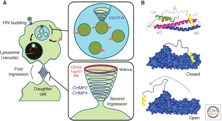
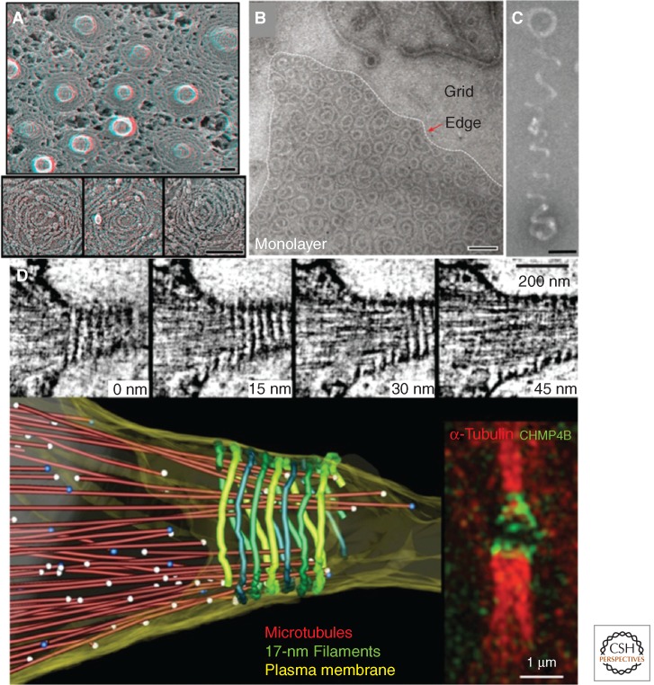
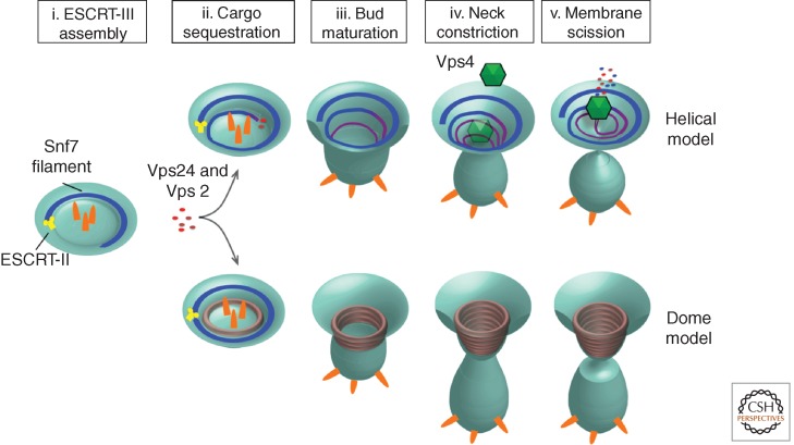
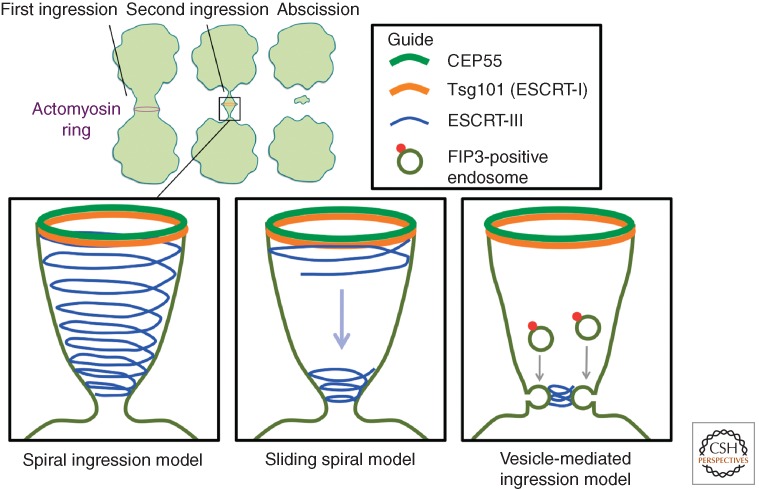
The endosomal sorting complexes required for transport (ESCRT) drive multivesicular body (MVB) biogenesis and cytokinetic abscission. Originally identified through genetics and cell biology, more recent work has begun to elucidate the molecular mechanisms of ESCRT-mediated membrane remodeling, with special focus on the ESCRT-III complex. In particular, several light and electron microscopic studies provide high-resolution imaging of ESCRT-III rings and spirals that purportedly drive MVB morphogenesis and abscission. These studies highlight unifying principles to ESCRT-III function, in particular: (1) the ordered assembly of the ESCRT-III monomers into a heteropolymer, (2) ESCRT-III as a dynamic complex, and (3) the role of the AAA ATPase Vps4 as a contributing factor in membrane scission. Mechanistic comparisons of ESCRT-III function in MVB morphogenesis and cytokinesis suggest common mechanisms in membrane remodeling.


Figures

Figure 1.
Figure 1.
ESCRT-mediated membrane remodeling in cellular physiology and pathophysiology. (A) Schematic of ESCRT-III helices driving MVB cargo sorting, HIV budding, and cell division. (B) Helical model of a Snf7 subunit from Saccharomyces cervisiae (top). ESCRT-III subunits undergo conformational changes from a closed (middle) to open state (bottom).
Figure 2.
Figure 2.
High-resolution imaging of ESCRT-III assemblies. (A) Electron micrograph of circular array protein scaffolds on membrane tubules in COS-7 cells expressing Snf7-1/CHMP4A and VPS4B(E235Q)-GFP. (Image from Hanson et al. 2008; adapted, with permission, from The Rockefeller University Press © 2008.) (B) Electron micrograph of reconstituted yeast Snf7 spirals assembled on lipid monolayers. (C) A helical polymer of reconstituted yeast Snf7, Vps24, and Vps2, imaged by electron microscopy. (Panels B and C are from Henne et al. 2012; adapted, with permission, from Cell Press © 2012.) (D) Electron tomography of high-pressure frozen cells, which show 17-nm filaments spiraling along the constriction zone. (This image is from Guizetti et al. 2011; adapted, with permission, from the author.) Scale bars, 100 nm, unless stated otherwise.
Figure 3.
Figure 3.
Helical and dome models for ESCRT-III-mediated membrane remodeling and vesicle scission.
Figure 4.
Figure 4.
Three models for ESCRT-III-mediated abscission.

References

    1. Agromayor M, Carlton JG, Phelan JP, Matthews DR, Carlin LM, Ameer-Beg S, Bowers K, Martin-Serrano J 2009. Essential role of hIST1 in cytokinesis. Mol Biol Cell 20: 1374–1387 - PMC - PubMed
    1. Ali N, Zhang L, Taylor S, Mironov A, Urbe S, Woodman P 2013. Recruitment of UBPY and ESCRT exchange drive HD-PTP-dependent sorting of EGFR to the MVB. Curr Biol 23: 453–461 - PubMed
    1. Babst M 2011. MVB vesicle formation: ESCRT-dependent, ESCRT-independent and everything in between. Curr Opin Cell Biol 23: 452–457 - PMC - PubMed
    1. Bajorek M, Schubert HL, McCullough J, Langelier C, Eckert DM, Stubblefield WM, Uter NT, Myszka DG, Hill CP, Sundquist WI 2009. Structural basis for ESCRT-III protein autoinhibition. Nat Struct Mol Biol 16: 754–762 - PMC - PubMed
    1. Bastos RN, Barr FA 2010. Plk1 negatively regulates Cep55 recruitment to the midbody to ensure orderly abscission. J Cell Biol 19: 751–760 - PMC - PubMed

Publication types

MeSH terms

Substances