Review
. 2013 Sep 1;5(9):a017905.
doi: 10.1101/cshperspect.a017905.

Nucleosome remodeling and epigenetics

Affiliations
Review

Nucleosome remodeling and epigenetics

Peter B Becker et al. Cold Spring Harb Perspect Biol. .

Abstract

Eukaryotic chromatin is kept flexible and dynamic to respond to environmental, metabolic, and developmental cues through the action of a family of so-called "nucleosome remodeling" ATPases. Consistent with their helicase ancestry, these enzymes experience conformation changes as they bind and hydrolyze ATP. At the same time they interact with DNA and histones, which alters histone-DNA interactions in target nucleosomes. Their action may lead to complete or partial disassembly of nucleosomes, the exchange of histones for variants, the assembly of nucleosomes, or the movement of histone octamers on DNA. "Remodeling" may render DNA sequences accessible to interacting proteins or, conversely, promote packing into tightly folded structures. Remodeling processes participate in every aspect of genome function. Remodeling activities are commonly integrated with other mechanisms such as histone modifications or RNA metabolism to assemble stable, epigenetic states.


Figures

Figure 1.
Figure 1.
DNase I-hypersensitivity (DH) analysis reveals rapid and reversible local nucleosome remodeling in vivo. The figure shows primary data from a classical DH analysis (Reik et al. 1991). The chromatin organization at the glucocorticoid-responsive enhancer element, 2.5 kb upstream of the promoter of the tyrosine aminotransferase gene, was probed in rat liver cells. Isolated nuclei of cells are digested with increasing amounts of DNase I. Digested genomic DNA is purified, cleaved with a restriction enzyme, resolved by agarose gel electrophoresis, and subjected to Southern blotting. The DH sites are revealed by indirect end-labeling of restriction fragments through hybridization of a small radioactive probe). They are marked with arrows. In the silent, uninduced state there are two DH sites at the promoter and one at −1 kb upstream. When the gene is activated on hormone induction with corticosterone, nucleosomes are remodeled at the enhancer within 15 min. A new DH site appears 2.5 kb upstream of the transcriptional start site, caused by chromatin remodeling (see “induced” columns). This correlates with the binding of glucocorticoid receptors and a complex set of remodeling factors. On removal of the hormone (“washout”), the factors dissociate and canonical nucleosomes reform within 15 min and the −2.5-kb enhancer DH disappears. The enhanced cleavage at the promoter reflects the transcriptional status of the gene.
Figure 2.
Figure 2.
Consequences of ATP-dependent nucleosome remodeling. (A) Models for nucleosome remodeling are illustrated by showing the change in position or composition of nucleosomes relative to the DNA wrapped around it. The left panel indicates a starting chromatin region with DNA reference points in the linker DNA or on the nucleosome shown in blue and pink, respectively. The right panels show possible outcomes of the remodeling reaction (from top to bottom): translational movement of a nucleosome (sliding) to expose a region that was previously occluded, exchange of a standard histone for a variant histone, and eviction of a nucleosome to expose the associated DNA. (B) Some nucleosome remodeling factors are also able to cooperate with histone chaperones to wrap DNA around histone octamers to generate nucleosomes. (C) Nucleosome remodeling factors may equilibrate the distances between nucleosomes in irregular arrays in a process termed nucleosome “spacing.”
Figure 3.
Figure 3.
The ISWI ATPase resides in several remodeling factors. The known ISWI-containing remodeling complexes in Drosophila are schematically shown. The functions of ACF, CHRAC, RSF, and NURF are described in the text. In mammals, the NoRC remodeler is involved in regulating the activity of ribosomal RNA genes (Li et al. 2006). NoRC is defined by the signature factor Tip5. The homologous protein in flies, toutatis, also interacts with ISWI (Vanolst et al. 2005). NoRC interacts with CtBP to form ToRC, which is involved in transcription regulation and nucleosome assembly outside of the nucleolus (Emelyanov et al. 2012). In mammals, additional complexes are known (Bao and Shen 2011; Kasten et al. 2011; Sims and Wade 2011; Yadon and Tsukiyama 2011) and it is likely that further assemblies will be discovered in flies as well.
Figure 4.
Figure 4.
Sequence relationships for the Snf2 family. Cladogram showing the relationship of the Snf2 family to other helicase-like proteins of superfamily 2 (SF2) (Fairman-Williams et al. 2010). Subfamily relationships within the Snf2 family are indicated based on alignments of the helicase-like region from 1306 members (Flaus et al. 2006). Branch lengths are not to scale. Swi2/Snf2 is the founding member of the Snf2 family of remodelers. (Adapted from Flaus et al. 2006.)
Figure 5.
Figure 5.
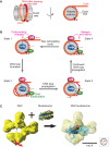
Mechanism of nucleosome remodeling. (A) Nucleosome view emphasizing the left hand wrapping of DNA. (Left) Side view of the nucleosome core with the histone octamer represented as a gray transparent cylinder and the DNA in orange (before the dyad axis) and red (after the dyad axis). (Right) Top view of the nucleosome (rotated 90º) in which the DNA after the dyad axis is represented by red dots. The star represents a reference point on the DNA sequence. (B) Model for DNA movement across the histone octamer during a remodeling event by the ISWI-type enzymes. Successive steps in a remodeling event are represented by States I–IV. In State I the DNA binding domain (DBD) is bound to the linker DNA and the translocase (Tr) domain is bound near the nucleosome dyad. A hypothetical “hinge” mediates the changes in conformation. In State II a conformational change between the DBD and the Tr “pulls in” DNA, which becomes visible as a bulge on the histone octamer surface. The Tr activity propagates this bulge across the surface of the histone octamer beyond the dyad axis (State III). The DNA loop continues to diffuse across the octamer surface and is released into the distal linker DNA (State IV). Loop diffusion thus effectively repositions the histone octamer relative to the DNA sequence (i.e., the star has moved closer to the dyad axis). A further conformational change triggered by aspects of the ATPase cycle lead to a resetting of the remodeler relative to the histone octamer (compare in States IV and I). The remodeler now engages with a different segment of linker DNA to start another cycle of remodeling. (A,B, Adapted from Clapier and Cairns 2009.) (C) Cryo-electron microscopy (EM) analysis of the RSC structure and nucleosome interaction. The yeast RSC resembles the Swi/Snf complex in subunit composition and overall architecture. Instead of the ATPase Swi2/Snf2, it contains the Sth1ATPase, which belongs to the same subfamily (Clapier and Cairns 2009). A 25-Å cryo-EM map of RSC (left) shows a central cavity that closely matches the shape and dimensions of a nucleosome core particle. Movement (indicated by the red arrow) of the bottom RSC domain appears to control access to the central cavity. Incubation of RSC with nucleosome core particles (NCPs) results in formation of a RSC-NCP complex (right panel) in which NCP density is apparent in the central RSC cavity. Interestingly, interaction with RSC in the absence of any ATP hydrolysis appears to result in extensive changes in NCP organization. Histone density can be identified, but nucleosomal DNA appears disordered (semitransparent blue density). This loosening of DNA may facilitate DNA translocation during remodeling. (Image and interpretation provided by Francisco Asturias, Scripps Research Institute, La Jolla.)
Figure 6.
Figure 6.
Four ATPase subfamilies: Signatures of SNF2, ISWI, CHD, and Ino80. The grouping of the remodeling ATPase of the Snf2 family is defined by signature motifs within the ATPase domain, whereas additional domains define the subfamilies. Members of the INO80 (and Swr1) subfamily of ATPases have a longer insertion between the two ATPase subdomains than other remodelers (Fig. 5). These subfamilies also contain a HSA (helicase-SANT) domain. The SWI/SNF family of ATPases contains a HSA domain, but is further defined by a carboxy-terminal bromodomain (capable of binding acetylated lysine residues). The ISWI and CHD family of ATPases each have SANT-SLIDE modules (blue) whereas only the CHD family has tandem chromodomains. (Adapted from Clapier and Cairns 2009.)
Figure 7.
Figure 7.
Example of a complex remodeling machine: INO80. The Saccharomyces cerevisiae INO80 complex provides an example of the subunit composition of a complex nucleosome-remodeling machine. INO80 subunits include the core ATPase, Ino80 (INOsitol requiring), Rvb1 (RuVB-like), Rvb2, Act1 (actin), Arp4 (actin-related protein), Arp5, Arp8, Nhp10 (nonhistone protein), Taf14 (TATA-binding protein-associated factor), Ies1 (Ino eighty subunit), Ies2, Ies3, Ies4, Ies5, and Ies6. (Adapted from Bao and Shen 2011.)
Figure 8.
Figure 8.
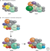
Diversity of Swi/Snf complexes in metazoa. (A) BAP and PBAP complexes in Drosophila. There are two distinct Swi/Snf-type complexes in Drosophila: the BRM-associated proteins complex (BAP) and the polybromo-containing BAP complex (PBAP). Although these complexes share multiple subunits including the ATPase BRM, they each have distinct subunits. The OSA, dD4, and TTH subunits are only found in the BAP complex and not in the PBAP complex. In contrast, Polybromo, BAF170, dBRD7, and SAYP are subunits of the PBAP complex but not the BAP complex. (Adapted from Ho and Crabtree 2010, with updates from Moshkin et al. 2012.) (B) Cell- and tissue-specific versions of BAF complexes in mammals. These complexes contain either the BRG1 or BRM ATPases. They may also contain polybromo (PB) and BAF200 (PBAF complexes) or BAF250A/B (BAF complexes). Shown here are composite representations of those variants. This figure serves to illustrate tissue-specific assemblies of BAF complexes, which have distinct functions in specific cell types. The subunits present in BAF complexes for each tissue are indicated (e.g., BAF60A or C). PB is colored the same as in A. The other subunits shaded with color are those whose presence varies in the different tissue, and define the tissue-specific complexes. (Adapted from Ho and Crabtree 2010.)
Figure 9.
Figure 9.
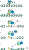
Model for the action of Swi/Snf-type nucleosome remodelers at promoters. and their regulation by acetylation. (A) SAGA or other histone acetyltransferase (HAT) complexes can be recruited to gene promoters by interacting with sequence-specific DNA-binding transcription activators (TA). Once recruited these HATs acetylate (blue Ac flag) nucleosomes in proximity of the activator recognition site. (B) The Swi/Snf or RSC nucleosome remodeling complexes (remodeler) can be recruited to promoters by interactions with transcription activators. Bromodomains (bromo) within subunits of these complexes then interact with the acetylated (Ac) nucleosomes at the promoter. (C) ATP-dependent remodeling and/or displacement (gray arrows) are preferentially directed at the acetylated nucleosomes bound by the bromodomains in the remodeling complex. (D) SAGA or other Gcn5-containing complexes acetylate specific lysines within subunits of the remodeling complex. These acetylated lysines compete for interaction with the bromodomains, which (E) leads to dissociation of the remodeler from the acetylated nucleosomes (gray arrow). (Adapted from Suganuma and Workman 2011.)

References

    1. Alkhatib SG, Landry JW 2011. The nucleosome remodeling factor. FEBS Lett 585: 3197–3207 - PMC - PubMed
    1. Almouzni G, Cedar H 2014. Maintenance of epigenetic information. Cold Spring Harb Perspect Biol 10.1101/cshperspect.a019372 - DOI - PMC - PubMed
    1. Altaf M, Auger A, Covic M, Cote J 2009. Connection between histone H2A variants and chromatin remodeling complexes. Biochem Cell Biol 87: 35–50 - PubMed
    1. Altaf M, Saksouk N, Cote J 2007. Histone modifications in response to DNA damage. Mutat Res 618: 81–90 - PubMed
    1. Bao Y, Shen X 2007. Chromatin remodeling in DNA double-strand break repair. Curr Opin Genet Dev 17: 126–131 - PubMed

Publication types

MeSH terms

LinkOut - more resources