. 2013 Oct 18;288(42):30270-30284.
doi: 10.1074/jbc.M113.510966. Epub 2013 Sep 3.

Arginine residues on the opposite side of the active site stimulate the catalysis of ribosome depurination by ricin A chain by interacting with the P-protein stalk

Affiliations

Arginine residues on the opposite side of the active site stimulate the catalysis of ribosome depurination by ricin A chain by interacting with the P-protein stalk

Xiao-Ping Li et al. J Biol Chem. .

Abstract

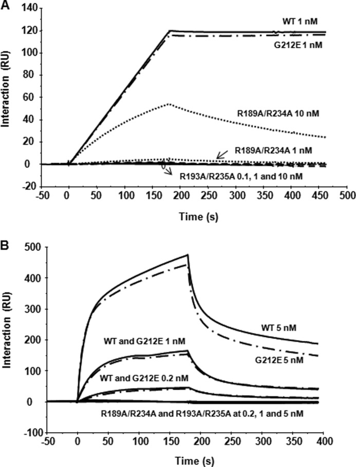
Ricin inhibits protein synthesis by depurinating the α-sarcin/ricin loop (SRL). Ricin holotoxin does not inhibit translation unless the disulfide bond between the A (RTA) and B (RTB) subunits is reduced. Ricin holotoxin did not bind ribosomes or depurinate them but could depurinate free RNA. When RTA is separated from RTB, arginine residues located at the interface are exposed to the solvent. Because this positively charged region, but not the active site, is blocked by RTB, we mutated arginine residues at or near the interface of RTB to determine if they are critical for ribosome binding. These variants were structurally similar to wild type RTA but could not bind ribosomes. Their K(m) values and catalytic rates (k(cat)) for an SRL mimic RNA were similar to those of wild type, indicating that their activity was not altered. However, they showed an up to 5-fold increase in K(m) and up to 38-fold decrease in kcat toward ribosomes. These results suggest that the stalk binding stimulates the catalysis of ribosome depurination by RTA. The mutated arginines have side chains behind the active site cleft, indicating that the ribosome binding surface of RTA is on the opposite side of the surface that interacts with the SRL. We propose that stalk binding stimulates the catalysis of ribosome depurination by orienting the active site of RTA toward the SRL and thereby allows docking of the target adenine into the active site. This model may apply to the translation factors that interact with the stalk.

Keywords: Protein Synthesis; RIP; Ribosomal RNA (rRNA); Ribosome-inactivating Protein; Ribosomes; Ricin; Toxins.


Figures

FIGURE 1.
FIGURE 1.
Interaction of RTA and ricin holotoxin with ribosomes and depurination of total RNA and ribosomes. A, RTA crystal structure (PDB code 1RTC) oriented to show residues at the active site and residues involved in SRL binding. RTA is shown in green. Arg180 (R180; orange), Glu177 (E177; red), and Gly212 (G212; blue) are shown to indicate the active site area. Residues predicted to be involved in binding the SRL are shown in cyan. B, ricin holotoxin crystal structure (PDB code 2AAI) in a similar orientation as in A, to show that the SRL binding site and the active site are not blocked by RTB (yellow). C, depurination of RNA by ricin holotoxin. Ricin holotoxin was incubated with yeast total RNA (1 μg) in the presence or absence of 1 mm TCEP, and depurination was quantified using qRT-PCR (47). D, depurination of yeast ribosomes by ricin holotoxin. Ribosomes (40 nm) were treated with ricin holotoxin in the presence or absence of 1 mm TCEP, RNA was extracted, and depurination was quantified using qRT-PCR (47). E, interaction of ricin holotoxin and RTA with yeast ribosomes by Biacore analysis. RTA (dashed lines) and ricin (solid lines) were immobilized on a CM5 chip of a Biacore T200 using amine coupling at 4234 and 7040 RU, respectively. EGFP was immobilized on the reference surface at 4588 RU as a control. The ribosomes at 0.25, 0.5, 1, 2, 4, 8, and 16 nm were passed over each surface at 40 μl/min for 5 min. Dissociation was at the same flow rate for another 5 min. The rate constants and the equilibrium dissociation constant for RTA are shown based on fitting of the data with the heterogeneous ligand parallel reaction model using Biacore T200 evaluation software. Error bars, S.D.
FIGURE 2.
FIGURE 2.
Viability, RTA expression, and depurination activity of RTA variants in yeast. A, ricin holotoxin (PDB code 2AAI) oriented to show RTA residues involved in ribosome binding (magenta). Arg196 and Arg197 do not interact with RTB (yellow), but the presence of RTB prevents ribosomal access to these amino acids. A few of the residues at the SRL binding site (cyan) are visible on the right. The rest of RTA is green. B, RTA (PDB code 1RTC) oriented to show ribosome-binding residues. Residues at the ribosome binding site are in magenta. Only some of the residues at the putative SRL binding site are visible and are shown in cyan. C, viability of yeast expressing RTA variants. Yeast cells were transformed with wild type RTA or RTA variants under the GAL1 promoter. Cells carrying the empty vector were used as controls. Yeast cells were first grown in SD medium supplemented with 2% glucose and then transferred to SD medium supplemented with 2% galactose. At 0, 4, 6, and 8 h postinduction, a series of 10-fold dilutions were plated on media containing 2% glucose and grown at 30 °C for 2–3 days. D, immunoblot analysis of RTA expression in yeast at 4 hpi. Total protein extracted from equal amount of cells (A600 of 0.8) was loaded on the gel, except only half the amount of cells (A600 of 0.4) was used for G212E due to the higher expression of this variant in yeast (40). Monoclonal antibodies against phosphoglycerate kinase 1 (Pgk1) and dolichoylphosphate mannosyltransferase 1 (Dpm1) were used as loading controls for cytosol and membranes, respectively. E, ribosome depurination in yeast expressing arginine variants of RTA. Total RNA (375 ng) was used to quantify the relative level of depurination using qRT-PCR by the comparative ΔCT method (ΔΔCT) (47). The -fold increase in depurination in yeast expressing RTA variants compared with yeast harboring the empty vector is shown. Error bars, S.D.
FIGURE 3.
FIGURE 3.
Partial digestion and far UV and near UV CD spectra of purified RTA variants. A, partial digestion of RTA variants with different proteinases. 3 μg of each RTA was digested with 0.15 μg of Glu-C (20:1) in 1× Glu-C buffer in a total volume of 10 μl at 25 °C for 5 h. 10 μg of RTA was digested with 0.4 μg of TPCK-treated trypsin (25:1) in 1× digestion buffer at 25 °C for 60 min. 10 μg of RTA was digested with 0.4 μg of TLCK-treated chymotrypsin (25:1) in buffer containing 100 mm Tris-HCl, pH 8.8, and 2 mm CaCl2 at 25 °C for 30 min. Shown are the far UV CD spectra (B) and near UV CD spectra (C) of RTA variants. The CD spectra of purified variants were determined at 25 °C in 50 mm phosphate buffer, pH 6.0, after overnight dialysis at 4 °C, followed by centrifugation or filtering. For the near-UV (250–350 nm), the concentrations ranged from 1.3 × 10⁻⁵ to 3.3 × 10⁻⁵ m depending on the variant, whereas for the far-UV, the concentrations were 2.69 × 10⁻⁶ to 4.09 × 10⁻⁶ m. Samples for CD were scanned eight times and averaged as described under “Experimental Procedures.”
FIGURE 4.
FIGURE 4.
Interaction of RTA variants with the ribosomal stalk pentamer and with monomeric ribosomes. A, N-terminal His10-tagged RTA variants were captured on an NTA chip at 2300 RU, and the same amount of EGFP was captured on the reference channel as control. Isolated stalk pentamer was passed over the RTA surface at 0.1, 1, and 10 nm as the analyte. B, RTA variants were captured on an NTA chip at 2000 RU, and the same amount of EGFP was captured to the reference channel as a control. Yeast ribosomes at a concentration of 0.2, 1, and 5 nm were passed over the surface as the analyte.
FIGURE 5.
FIGURE 5.
Depurination of ribosomes and total RNA by RTA variants in vitro. A, yeast ribosomes (40 nm) were incubated with different concentrations of RTA, varying from 0 to 40 nm. The extent of depurination was quantified by qRT-PCR as described previously (47). RTA concentrations are shown in log scale due to large differences in the activities of wild type RTA and arginine variants. The y axis shows the -fold change in depurination relative to the control samples without toxin treatment. B, total RNA (1 μg) was treated with different concentrations of RTA as described under “Experimental Procedures.” The extent of depurination was quantified by qRT-PCR as described previously (47). Error bars, S.D.
FIGURE 6.
FIGURE 6.
Kinetic curve fits for catalysis of depurination of ribosomes and stem-loop RNA by RTA variants. A, Michaelis-Menten fits of ribosome depurination performed with the continuous assay described under “Experimental Procedures.” Wild type RTA was used at 1 nm, R189A/R234A at 10 nm, G212E at 30 nm, and R193A/R235A at 50 nm, all with increasing amounts of ribosomes as indicated. The RTA-independent rate of adenine generation was subtracted for each. Adenine standards covering the range of depurination were measured for each mutant. B, Michaelis-Menten fits of depurination of 10-mer loop performed with the discontinuous assay at pH 4.5 described under “Experimental Procedures.” Wild type RTA was used at 50 nm, R189A/R234A at 50 nm, G212E at 100 nm, and R193A/R235A at 50 nm. Adenine standards were measured for each under the same buffer conditions. Results from a representative experiment are shown.
FIGURE 7.
FIGURE 7.
Model showing how RTA accesses the SRL. S. cerevisiae 26 S rRNA (PDB code 3U5H) and 60 S subunit (PDB code 3U5I) are indicated as light gray and dark gray colors, respectively. The fitted schematic structure of P0 fragment complexed with the N-terminal domain of P-proteins (PDB code 3A1Y) from Archaea is depicted as yellow and green, respectively. One CTD domain of a P-protein is shown as a gray line attached to the RTA molecule. The yeast 60 S subunit is oriented to show A3027 (red) of the SRL (cyan) and RTA (PDB code 1RTC) oriented to show R180 (orange) at the active site, the putative SRL binding site (cyan), and the stalk binding site (magenta). In step 1, RTA binds to the C-terminal tail of a P-protein using arginine residues located on the stalk binding surface. In step 2, the flexible hinge of the P-protein orients the active site of RTA toward the SRL and properly positions A3027 of the SRL into the active site of RTA. This mechanism enables RTA to establish the specific contacts with the backbone of the SRL necessary to hydrolyze the N-glycosidic bond of A3027. An enlarged image of the interaction is shown in the inset.

Similar articles

Cited by

References

    1. Audi J., Belson M., Patel M., Schier J., Osterloh J. (2005) Ricin poisoning. A comprehensive review. JAMA 294, 2342–2351 - PubMed
    1. Spooner R. A., Watson P. D., Marsden C. J., Smith D. C., Moore K. A., Cook J. P., Lord J. M., Roberts L. M. (2004) Protein disulphide-isomerase reduces ricin to its A and B chains in the endoplasmic reticulum. Biochem. J. 383, 285–293 - PMC - PubMed
    1. Endo Y., Mitsui K., Motizuki M., Tsurugi K. (1987) The mechanism of action of ricin and related toxic lectins on eukaryotic ribosomes. The site and the characteristics of the modification in 28 S ribosomal RNA caused by the toxins. J. Biol. Chem. 262, 5908–5912 - PubMed
    1. Montanaro L., Sperti S., Stirpe F. (1973) Inhibition by ricin of protein synthesis in vitro. Ribosomes as the target of the toxin. Biochem. J. 136, 677–683 - PMC - PubMed
    1. Olsnes S., Refsnes K., Pihl A. (1974) Mechanism of action of the toxic lectins abrin and ricin. Nature 249, 627–631 - PubMed

Publication types

LinkOut - more resources