. 2013 Oct 18;288(42):30544-30557.
doi: 10.1074/jbc.M113.495465. Epub 2013 Sep 3.

Endogenous interleukin-1β in neuropathic rats enhances glutamate release from the primary afferents in the spinal dorsal horn through coupling with presynaptic N-methyl-D-aspartic acid receptors

Affiliations

Endogenous interleukin-1β in neuropathic rats enhances glutamate release from the primary afferents in the spinal dorsal horn through coupling with presynaptic N-methyl-D-aspartic acid receptors

Xisheng Yan et al. J Biol Chem. .

Abstract

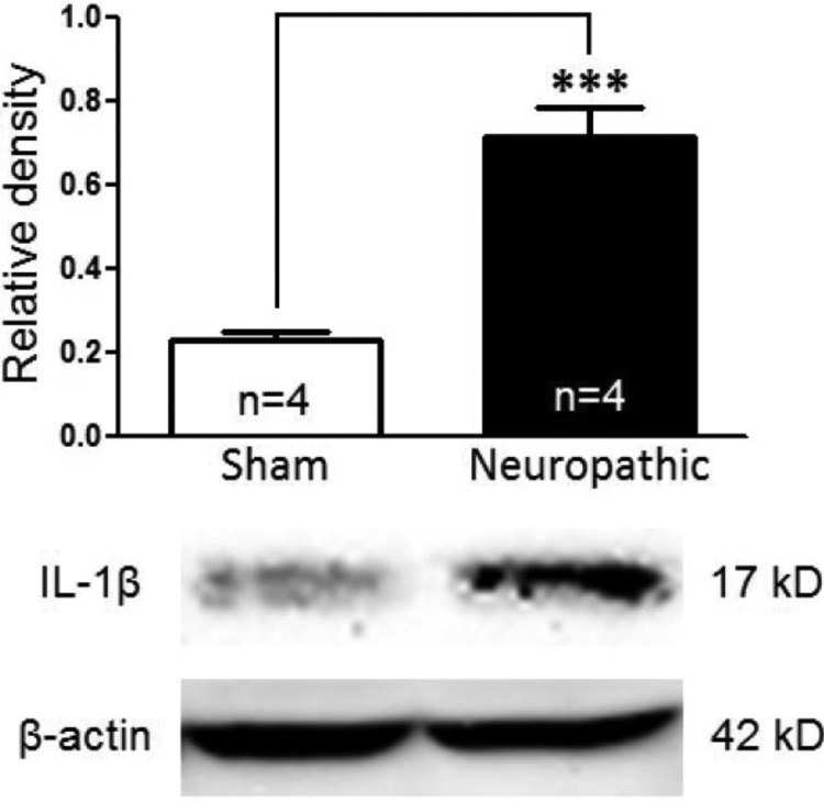
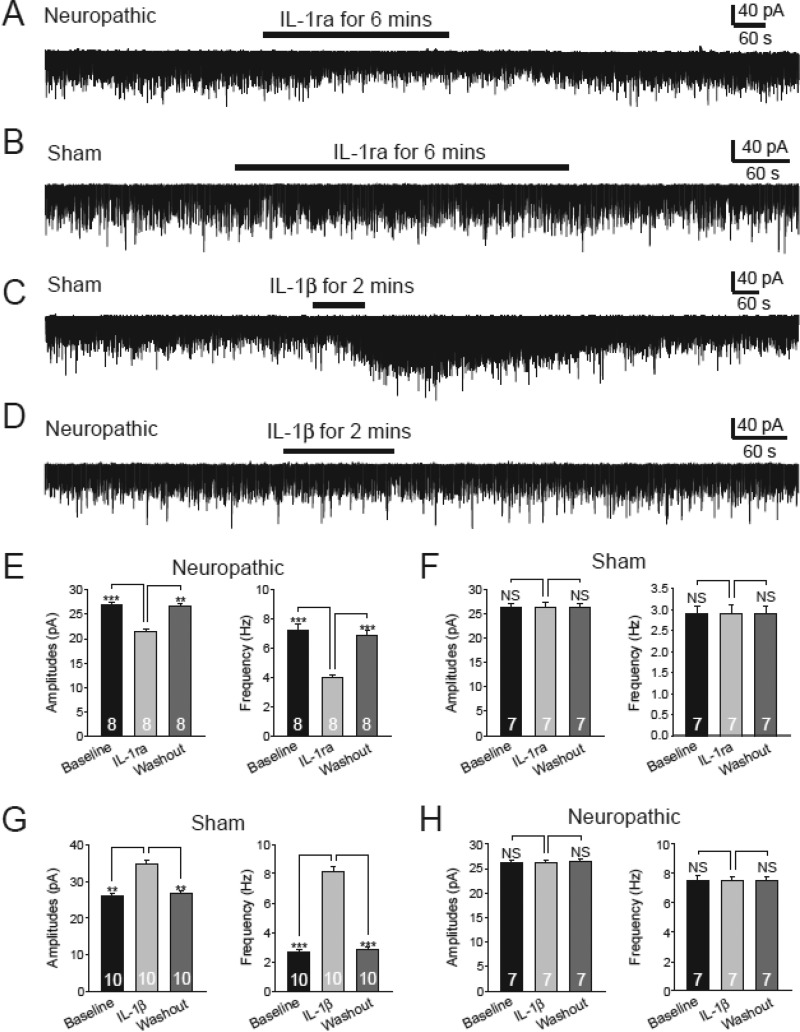
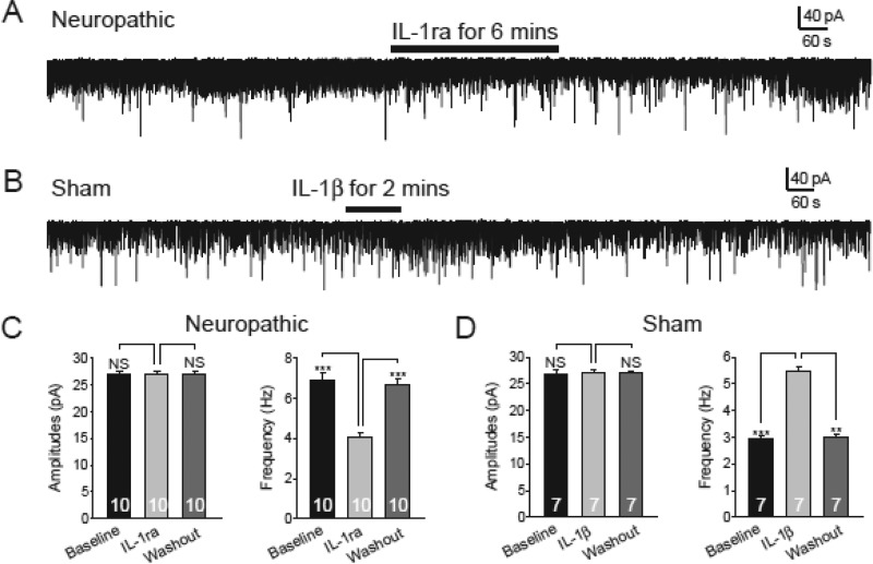
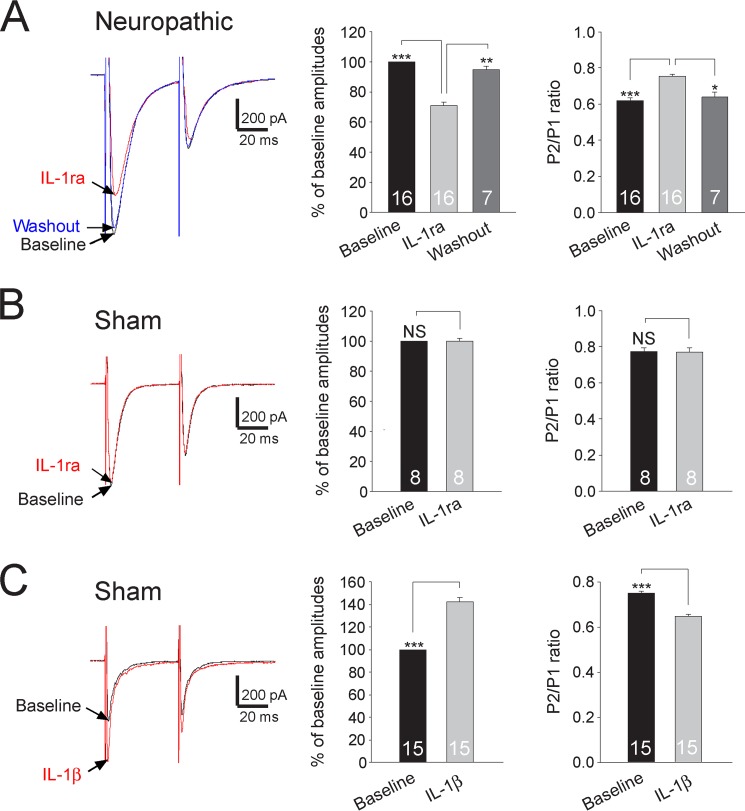
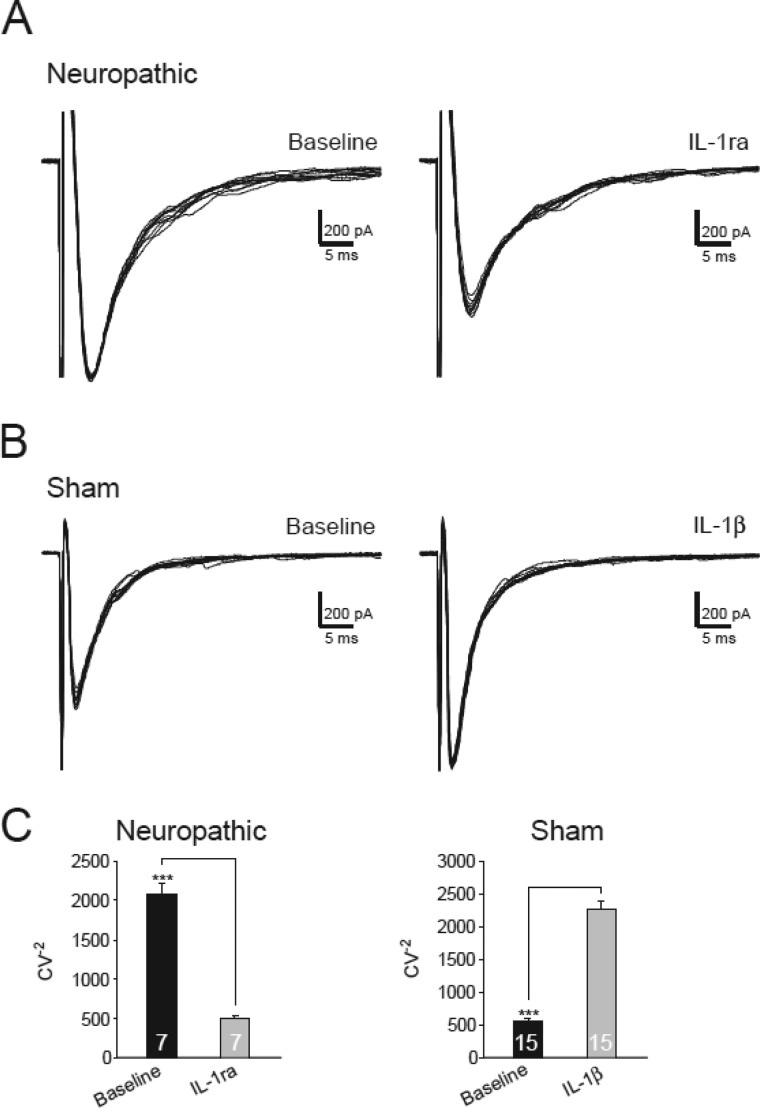
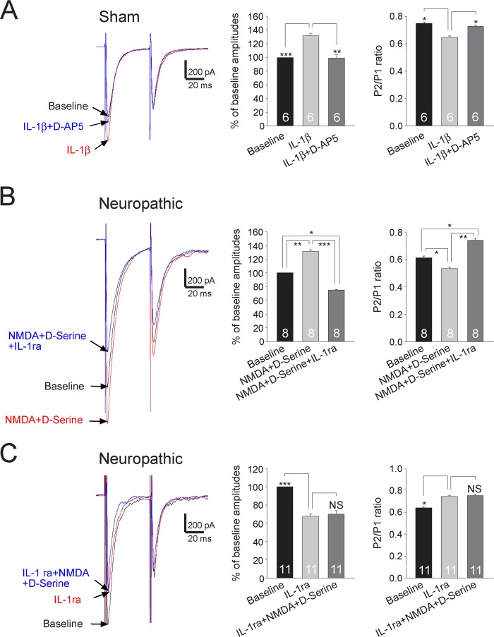
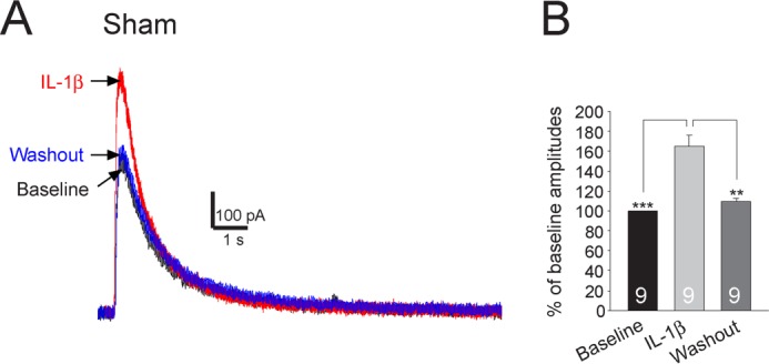
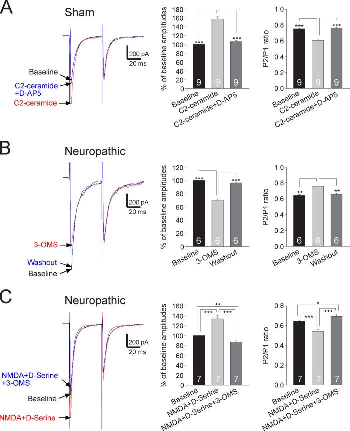
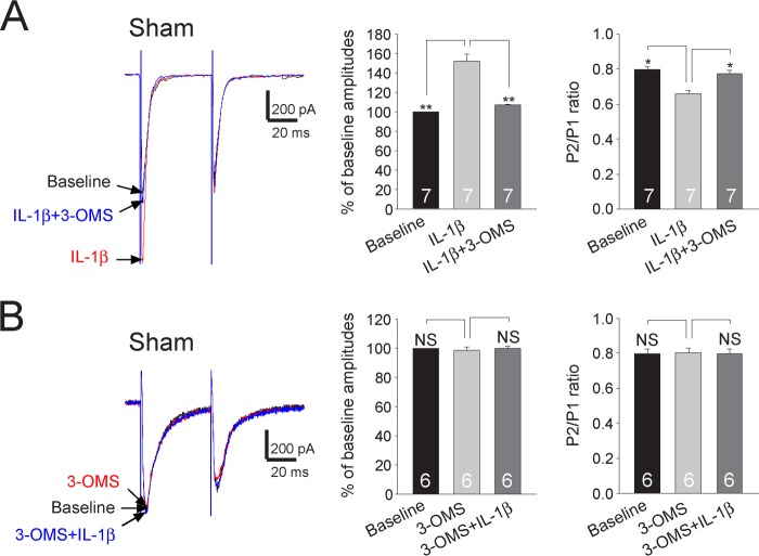
Excessive activation of glutamate receptors and overproduction of proinflammatory cytokines, including interleukin-1β (IL-1β) in the spinal dorsal horn, are key mechanisms underlying the development and maintenance of neuropathic pain. In this study, we investigated the mechanisms by which endogenous IL-1β alters glutamatergic synaptic transmission in the spinal dorsal horn in rats with neuropathic pain induced by ligation of the L5 spinal nerve. We demonstrated that endogenous IL-1β in neuropathic rats enhances glutamate release from the primary afferent terminals and non-NMDA glutamate receptor activities in postsynaptic neurons in the spinal dorsal horn. Myeloid differentiation primary response protein 88 (MyD88) is a mediator used by IL-1β to enhance non-NMDA glutamate receptor activities in postsynaptic neurons in the spinal dorsal horn. Presynaptic NMDA receptors are effector receptors used by the endogenous IL-1β to enhance glutamate release from the primary afferents in neuropathic rats. This is further supported by the fact that NMDA currents recorded from small neurons in the dorsal root ganglion of normal rats are potentiated by exogenous IL-1β. Furthermore, we provided evidence that functional coupling between IL-1β receptors and presynaptic NMDA receptors at the primary afferent terminals is mediated by the neutral sphingomyelinase/ceramide signaling pathway. Hence, functional coupling between IL-1β receptors and presynaptic NMDA receptors at the primary afferent terminals is a crucial mechanism leading to enhanced glutamate release and activation of non-NMDA receptors in the spinal dorsal horn neurons in neuropathic pain conditions. Interruption of such functional coupling could be an effective approach for the treatment of neuropathic pain.

Keywords: Cell Signaling; Glia; Glutamate Receptors Ionotropic (AMPA, NMDA); Pain; Patch Clamp.


Figures

FIGURE 1.
FIGURE 1.
Protein expression of IL-1β in the spinal dorsal horn ipsilateral to the injury site in neuropathic rats is increased. Samples of IL-1β expression in the spinal dorsal horn at the L4 to L5 segment in neuropathic (n = 4) and sham-operated (n = 4) rats are shown. The bar graph shows the mean (+S.E.) relative density to β-actin in each group. ***, p < 0.001.
FIGURE 2.
FIGURE 2.
Endogenous IL-1β in neuropathic rats enhances glutamate release from presynaptic terminals and activities of non-NMDA glutamate receptors in dorsal horn postsynaptic neurons in neuropathic rats. Inhibition of IL-1β receptors with bath application of IL-1ra (100 ng/ml) significantly and reversibly reduced both the frequency and amplitudes of mEPSCs in neuropathic rats (A and E) but had no effects on mEPSCs in sham-operated rats (B and F). Bath perfusion of IL-1β enhanced both the frequency and amplitudes of mEPSCs in sham-operated rats (C and G) but had no effects on mEPSCs in neuropathic rats (D and H). Bar graphs show the mean (+S.E.) amplitude and frequency for each tested agent. The number of neurons included in each group for the analysis is shown in each bar. **, p < 0.01; ***, p < 0.001; NS, no statistical significance.
FIGURE 3.
FIGURE 3.
MyD88 is a mediator used by IL-1β to alter non-NMDA glutamate receptor activities in the spinal dorsal horn neurons. Recordings were obtained with electrodes containing the MyD88 inhibitory peptide (1 mm) in the intracellular solution. Under this condition, bath perfusion of IL-1ra (100 ng/ml) reversibly and significantly reduced the mEPSC frequency but not the mEPSC amplitude in spinal slices of neuropathic rats (A and C). Bath perfusion of IL-1β (10 ng/ml) significantly increased the mEPSC frequency but not the mEPSC amplitude in spinal slices of sham-operated rats (B and D). Bar graphs show the mean (+S.E.) amplitude and frequency for each tested agent. The number of neurons included in each group for the analysis is shown in each bar. **, p < 0.01; ***, p < 0.001; NS, no statistical significance.
FIGURE 4.
FIGURE 4.
Endogenous IL-1β in neuropathic rats increases glutamate release from the primary afferent terminals in neuropathic rats. Samples of EPSCs evoked by a pair of electrical pulses applied to the spinal dorsal root before (base line) and after application of IL-1ra (100 ng/ml) recorded from neuropathic rats (A) and sham-operated rats (B). C, samples of EPSCs evoked by a pair of electrical pulses applied to the spinal dorsal root recorded from sham-operated rats at base line and after bath application of IL-1β (10 ng/ml). Bar graphs show the mean percentage (+ S.E.) of base line amplitude and mean (+ S.E.) P2/P1 ratios for each tested agent. The number of neurons included in each group for the analysis is shown in each bar. *, p < 0.05; **, p < 0.01; ***, p < 0.001; NS, no statistical significance.
FIGURE 5.
FIGURE 5.
Activation of IL-1β receptors increases the inverse squared coefficient of variation. A, samples of variability of 10 evoked EPSCs collected from a neuron from neuropathic rats before and after bath perfusion of IL-1ra (100 ng/ml). B, samples of variability of 10 evoked EPSCs collected from a neuron from sham-operated rats before and after bath perfusion of IL-1β (10 ng/ml). C, bar graphs show the mean (+ S.E.) CV−2 before (base line) and after perfusion of each tested agent. The number of neurons included in each group for the analysis is shown in each bar. ***, p < 0.001.
FIGURE 6.
FIGURE 6.
Presynaptic NMDA receptors are effector receptors used by the endogenous IL-1β to enhance glutamate release from the primary afferents in neuropathic rats. A, samples of EPSCs evoked by a pair of electrical pulses recorded from a neuron of sham-operated rats at base line, during perfusion of IL-1β (10 ng/ml), and then during addition of the NMDA receptor inhibitor D-AP5 (25 μm) in the presence of IL-1β. B, samples of EPSCs evoked by a pair of electrical pulses recorded from a neuron from neuropathic rats at base line, during perfusion of NMDA (50 μm) plus d-serine (200 μm), and then during addition of IL-1ra (100 ng/ml) in the presence of NMDA plus d-serine. C, samples of EPSCs evoked by a pair of electrical pulses recorded from a neuron from neuropathic rats at base line, during perfusion of IL-1ra (100 ng/ml), and then during addition of NMDA (50 μm) plus d-serine (200 μm) in the presence of IL-1ra. Bar graphs (right) show the mean percentage (+ S.E.) of base-line amplitude and mean (+ S.E.) P2/P1 ratios for the tested agents. The number of neurons included in each group for the analysis is shown in each bar. *, p < 0.05; **, p < 0.01; ***, p < 0.001. NS, no statistical significance.
FIGURE 7.
FIGURE 7.
NMDA currents recorded from small neurons in the dorsal root ganglion are significantly enhanced by IL-1β. Recordings of NMDA currents evoked by NMDA (50 μm) injected onto the recorded neuron by a puff electrode at base line, during, and after washout of IL-1β are shown in A. The mean (+S.E.) amplitudes of NMDA currents at base line, during, and after washout of IL-1β are shown in bar graphs (B). The number of neurons included for the analysis is shown in each bar. **, p < 0.01; ***, p < 0.001.
FIGURE 8.
FIGURE 8.
Presynaptic NMDA receptors are effector receptors used by the sphingomyelinase/ceramide pathway to enhance glutamate release from the primary afferents in neuropathic rats. A, samples of EPSCs evoked by a pair of electrical pulses recorded from a neuron of sham-operated rats at base line, during perfusion of C2-ceramide (2 μg/ml), and then during addition of the NMDA receptor inhibitor D-AP5 (25 μm) in the presence of C2-ceramide. B, samples of EPSCs evoked by a pair of electrical pulses recorded from a neuron of neuropathic rats at base line, during, and after washout of the neutral sphingomyelinase inhibitor (3-OMS, 30 μm). C, samples of EPSCs evoked by a pair of electrical pulses recorded from a neuron of neuropathic rats at base line, during perfusion of NMDA (50 μm) plus d-serine (200 μm), and then during addition of 3-OMS (30 μm) in the presence of NMDA plus d-serine. Bar graphs (right) show the mean percentage (+S.E.) of base-line amplitude and mean (+ S.E.) P2/P1 ratios for the tested agents. The number of neurons included in each group for the analysis is shown in each bar. *, p < 0.05; **, p < 0.01; ***, p < 0.001.
FIGURE 9.
FIGURE 9.
Enhancement of glutamate release from the primary afferent terminals by IL-1β is mediated by the sphingomyelinase/ceramide pathway. A, samples of EPSCs evoked by a pair of electrical pulses recorded from a neuron of sham-operated rats at base line, during perfusion of IL-1β (10 ng/ml), and then during addition of the neutral sphingomyelinase inhibitor (3-OMS, 30 μm) in the presence of IL-1β. B, samples of EPSCs evoked by a pair of electrical pulses recorded from a neuron of sham-operated rats at base line, during perfusion of 3-OMS (30 μm), and then during addition of IL-1β (10 ng/ml) in the presence of 3-OMS. Bar graphs (right) show the mean percentage (+S.E.) of base-line amplitude and mean (+S.E.) P2/P1 ratios for the tested agents. The number of neurons included in each group for the analysis is shown in each bar. *, p < 0.05; **, p < 0.01. NS, no statistical significance.

References

    1. Ren K., Dubner R. (2010) Interactions between the immune and nervous systems in pain. Nat. Med. 16, 1267–1276 - PMC - PubMed
    1. Milligan E. D., Watkins L. R. (2009) Pathological and protective roles of glia in chronic pain. Nat. Rev. Neurosci. 10, 23–36 - PMC - PubMed
    1. De Biasi S., Rustioni A. (1988) Glutamate and substance P coexist in primary afferent terminals in the superficial laminae of spinal cord. Proc. Natl. Acad. Sci. U.S.A. 85, 7820–7824 - PMC - PubMed
    1. Willis W. D. (2002) Long-term potentiation in spinothalamic neurons. Brain Res. Brain Res. Rev. 40, 202–214 - PubMed
    1. Danbolt N. C. (2001) Glutamate uptake. Prog. Neurobiol. 65, 1–105 - PubMed

Publication types

LinkOut - more resources