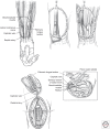
Primate models in organ transplantation
- PMID: 24003248
- PMCID: PMC3753726
- DOI: 10.1101/cshperspect.a015503
Primate models in organ transplantation
Abstract
Large animal models have long served as the proving grounds for advances in transplantation, bridging the gap between inbred mouse experimentation and human clinical trials. Although a variety of species have been and continue to be used, the emergence of highly targeted biologic- and antibody-based therapies has required models to have a high degree of homology with humans. Thus, the nonhuman primate has become the model of choice in many settings. This article will provide an overview of nonhuman primate models of transplantation. Issues of primate genetics and care will be introduced, and a brief overview of technical aspects for various transplant models will be discussed. Finally, several prominent immunosuppressive and tolerance strategies used in primates will be reviewed.
Figures


References
-
- Adams AB, Shirasugi N, Durham MM, Strobert E, Anderson D, Rees P, Cowan S, Xu H, Blinder Y, Cheung M, et al. 2002. Calcineurin inhibitor-free CD28 blockade-based protocol protects allogeneic islets in nonhuman primates. Diabetes 51: 265–270 - PubMed
-
- Adams AB, Pearson TC, Larsen CP 2003. Heterologous immunity: An overlooked barrier to tolerance. Immunol Rev 196: 147–160 - PubMed
-
- Adams AB, Shirasugi N, Jones TR, Durham MM, Strobert EA, Cowan S, Rees P, Hendrix R, Price K, Kenyon NS, et al. 2005. Development of a chimeric anti-CD40 monoclonal antibody that synergizes with LEA29Y to prolong iset allograft survival. J Immunol 174: 542–550 - PubMed
-
- Akalin E, Chandraker A, Russell ME, Turka LA, Hancock WW, Sayegh MH 1996. CD28-B7 T cell costimulatory blockade by CTLA4Ig in the rat renal allograft model: Inhibition of cell-mediated and humoral immune responses in vivo. Transplantation 62: 1942–1945 - PubMed
-
- Andrade MCR, Penedo MCT, Ward T, Silva VF, Bertolini LR, Roberts JA, Leite JP, Cabello PH 2004. Determination of genetic status in a closed colony of rhesus monkeys (Macaca mulatta). Primates 45: 183–186 - PubMed
Publication types
MeSH terms
Substances
LinkOut - more resources
Full Text Sources
Other Literature Sources
Medical
Miscellaneous
