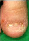
Onychomycosis in children: an experience of 59 cases
- PMID: 24003276
- PMCID: PMC3756198
- DOI: 10.5021/ad.2013.25.3.327
Onychomycosis in children: an experience of 59 cases
Abstract
Background: Although tinea unguium in children has been studied in the past, no specific etiological agents of onychomycosis in children has been reported in Korea.
Objective: The purpose of this study was to investigate onychomycosis in Korean children.
Methods: We reviewed fifty nine patients with onychomycosis in children (0~18 years of age) who presented during the ten-year period between 1999 and 2009. Etiological agents were identified by cultures on Sabouraud's dextrose agar with and without cycloheximide. An isolated colony of yeasts was considered as pathogens if the same fungal element was identified at initial direct microscopy and in specimen-yielding cultures at a follow-up visit.
Results: Onychomycosis in children represented 2.3% of all onychomycosis. Of the 59 pediatric patients with onychomycosis, 66.1% had toenail onychomycosis with the rest (33.9%) having fingernail onychomycosis. The male-to-female ratio was 1.95:1. Fourteen (23.7%) children had concomitant tinea pedis infection, and tinea pedis or onychomycosis was also found in eight of the parents (13.6%). Distal and lateral subungual onychomycosis was the most common (62.7%) clinical type. In toenails, Trichophyton rubrum was the most common etiological agent (51.3%), followed by Candida albicans (10.2%), C. parapsilosis (5.1%), C. tropicalis (2.6%), and C. guilliermondii (2.6%). In fingernails, C. albicans was the most common isolated pathogen (50.0%), followed by T. rubrum (10.0%), C. parapsilosis (10.0%), and C. glabrata (5.0%).
Conclusion: Because of the increase in pediatric onychomycosis, we suggest the need for a careful mycological examination of children who are diagnosed with onychomycosis.
Keywords: Child; Onychomycosis.
Figures


References
-
- Verma S, Heffernan MP. Superficial fungal infections: dermatophytosis, onychomycosis, tinea nigra, piedra. In: Wolff K, Goldsmith LA, Katz SI, Gilchrest BA, Paller AS, Leffell DJ, editors. Fitzpatrick's dermatology in general medicine. 7th ed. New York: McGraw-Hill; 2008. pp. 1807–1821.
-
- Chang P, Logemann H. Onychomycosis in children. Int J Dermatol. 1994;33:550–551. - PubMed
-
- Ploysangam T, Lucky AW. Childhood white superficial onychomycosis caused by Trichophyton rubrum: report of seven cases and review of the literature. J Am Acad Dermatol. 1997;36:29–32. - PubMed
-
- Gupta AK, Sibbald RG, Lynde CW, Hull PR, Prussick R, Shear NH, et al. Onychomycosis in children: prevalence and treatment strategies. J Am Acad Dermatol. 1997;36:395–402. - PubMed
-
- Lateur N, Mortaki A, André J. Two hundred ninety-six cases of onychomycosis in children and teenagers: a 10-year laboratory survey. Pediatr Dermatol. 2003;20:385–388. - PubMed
LinkOut - more resources
Full Text Sources
Other Literature Sources
Miscellaneous

