

3.0 Tesla vs 1.5 Tesla breast magnetic resonance imaging in newly diagnosed breast cancer patients
- PMID: 24003354
- PMCID: PMC3758496
- DOI: 10.4329/wjr.v5.i8.285
3.0 Tesla vs 1.5 Tesla breast magnetic resonance imaging in newly diagnosed breast cancer patients
Abstract
Aim: To compare 3.0 Tesla (T) vs 1.5T magnetic resonance (MR) imaging systems in newly diagnosed breast cancer patients.
Methods: Upon Institutional Review Board approval, a Health Insurance Portability and Accountability Act-compliant retrospective review of 147 consecutive 3.0T MR examinations and 98 consecutive 1.5T MR examinations in patients with newly diagnosed breast cancer between 7/2009 and 5/2010 was performed. Eleven patients who underwent neoadjuvant chemotherapy in the 3.0T group were excluded. Mammographically occult suspicious lesions (BIRADS Code 4 and 5) additional to the index cancer in the ipsilateral and contralateral breast were identified. Lesion characteristics and pathologic diagnoses were recorded, and results achieved with both systems compared. Statistical significance was analyzed using Fisher's exact test.
Results: In the 3.0T group, 206 suspicious lesions were identified in 55% (75/136) of patients and 96% (198/206) of these lesions were biopsied. In the 1.5T group, 98 suspicious lesions were identified in 53% (52/98) of patients and 90% (88/98) of these lesions were biopsied. Biopsy results yielded additional malignancies in 24% of patients in the 3.0T group vs 14% of patients in the 1.5T group (33/136 vs 14/98, P = 0.07). Average size and histology of the additional cancers was comparable. Of patients who had a suspicious MR imaging study, additional cancers were found in 44% of patients in the 3.0T group vs 27% in the 1.5T group (33/75 vs 14/52, P = 0.06), yielding a higher positive predictive value (PPV) for biopsies performed with the 3.0T system.
Conclusion: 3.0T MR imaging detected more additional malignancies in patients with newly diagnosed breast cancer and yielded a higher PPV for biopsies performed with the 3.0T system.
Keywords: 3 Tesla; Breast; Breast cancer; Breast magnetic resonance imaging; Cancer staging; Magnetic resonance imaging; Outcome; Technical.
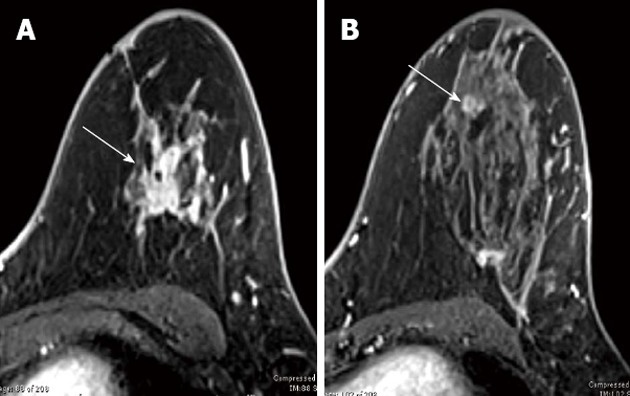
Figures

References
-
- Hecht EM, Lee RF, Taouli B, Sodickson DK. Perspectives on body MR imaging at ultrahigh field. Magn Reson Imaging Clin N Am. 2007;15:449–465, viii. - PubMed
-
- Kuhl CK. Breast MR imaging at 3T. Magn Reson Imaging Clin N Am. 2007;15:315–320, vi. - PubMed
-
- Meeuwis C, Mann RM, Mus RD, Winkel A, Boetes C, Barentsz JO, Veltman J. MRI-guided breast biopsy at 3T using a dedicated large core biopsy set: feasibility and initial results. Eur J Radiol. 2011;79:257–261. - PubMed
-
- Soher BJ, Dale BM, Merkle EM. A review of MR physics: 3T versus 1.5T. Magn Reson Imaging Clin N Am. 2007;15:277–290, v. - PubMed
-
- Chatterji M, Mercado CL, Moy L. Optimizing 1.5-Tesla and 3-Tesla dynamic contrast-enhanced magnetic resonance imaging of the breasts. Magn Reson Imaging Clin N Am. 2010;18:207–224, viii. - PubMed
LinkOut - more resources
Full Text Sources
Other Literature Sources
