

Synergistic effect of Indian hedgehog and bone morphogenetic protein-2 gene transfer to increase the osteogenic potential of human mesenchymal stem cells
- PMID: 24004723
- PMCID: PMC3854715
- DOI: 10.1186/scrt316
Synergistic effect of Indian hedgehog and bone morphogenetic protein-2 gene transfer to increase the osteogenic potential of human mesenchymal stem cells
Abstract
Introduction: To stimulate healing of large bone defects research has concentrated on the application of mesenchymal stem cells (MSCs).
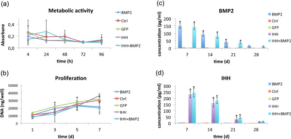
Methods: In the present study, we induced the overexpression of the growth factors bone morphogenetic protein 2 (BMP-2) and/or Indian hedgehog (IHH) in human MSCs by adenoviral transduction to increase their osteogenic potential. GFP and nontransduced MSCs served as controls. The influence of the respective genetic modification on cell metabolic activity, proliferation, alkaline phosphatase (ALP) activity, mineralization in cell culture, and osteogenic marker gene expression was investigated.
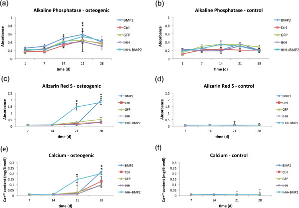
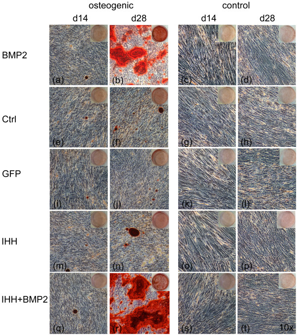
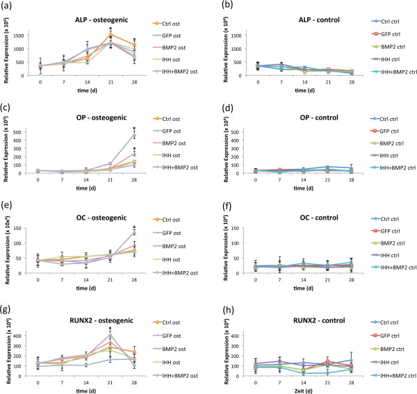
Results: Transduction had no negative influence on cell metabolic activity or proliferation. ALP activity showed a typical rise-and-fall pattern with a maximal activity at day 14 and 21 after osteogenic induction. Enzyme activity was significantly higher in groups cultured with osteogenic media. The overexpression of BMP-2 and especially IHH + BMP-2 resulted in a significantly higher mineralization after 28 days. This was in line with obtained quantitative reverse transcriptase polymerase chain reaction (qRT-PCR) analyses, which showed a significant increase in osteopontin and osteocalcin expression for osteogenically induced BMP-2 and IHH + BMP-2 transduced cells when compared with the other groups. Moreover, an increase in runx2 expression was observed in all osteogenic groups toward day 21. It was again more pronounced for BMP-2 and IHH + BMP-2 transduced cells cultured in osteogenic media.
Conclusions: In summary, viral transduction did not negatively influence cell metabolic activity and proliferation. The overexpression of BMP-2 in combination with or without IHH resulted in an increased deposition of mineralized extracellular matrix, and expression of osteogenic marker genes. Viral transduction therefore represents a promising means to increase the osteogenic potential of MSCs and the combination of different transgenes may result in synergistic effects.
Figures
Similar articles
-
Cx43- and Smad-Mediated TGF-β/ BMP Signaling Pathway Promotes Cartilage Differentiation of Bone Marrow Mesenchymal Stem Cells and Inhibits Osteoblast Differentiation.Cell Physiol Biochem. 2017;42(4):1277-1293. doi: 10.1159/000478957. Epub 2017 Jul 11. Cell Physiol Biochem. 2017. Retraction in: Cell Physiol Biochem. 2021;55(4):517. doi: 10.33594/000000409. PMID: 28697500 Retracted.
-
Pilose antler aqueous extract promotes the proliferation and osteogenic differentiation of bone marrow mesenchymal stem cells by stimulating the BMP-2/Smad1, 5/Runx2 signaling pathway.Chin J Nat Med. 2019 Oct;17(10):756-767. doi: 10.1016/S1875-5364(19)30092-5. Chin J Nat Med. 2019. PMID: 31703756
-
Noggin suppression decreases BMP-2-induced osteogenesis of human bone marrow-derived mesenchymal stem cells in vitro.J Cell Biochem. 2012 Dec;113(12):3672-80. doi: 10.1002/jcb.24240. J Cell Biochem. 2012. PMID: 22740073
-
The effect of five proteins on stem cells used for osteoblast differentiation and proliferation: a current review of the literature.Cell Mol Life Sci. 2014 Jan;71(1):113-42. doi: 10.1007/s00018-013-1326-0. Epub 2013 Apr 9. Cell Mol Life Sci. 2014. PMID: 23568025 Free PMC article. Review.
-
Evaluating Bioassays for the Determination of Simvastatin's Osteogenic Activity: A Systematic Review.J Funct Biomater. 2025 Feb 11;16(2):61. doi: 10.3390/jfb16020061. J Funct Biomater. 2025. PMID: 39997596 Free PMC article. Review.
Cited by
-
Research progress on the hedgehog signalling pathway in regulating bone formation and homeostasis.Cell Prolif. 2022 Jan;55(1):e13162. doi: 10.1111/cpr.13162. Epub 2021 Dec 16. Cell Prolif. 2022. PMID: 34918401 Free PMC article. Review.
-
Tendon-derived stem cells from the long head of the biceps tendon: Inflammation does not affect the regenerative potential.Bone Joint Res. 2019 Oct 3;8(9):414-424. doi: 10.1302/2046-3758.89.BJR-2018-0214.R2. eCollection 2019 Sep. Bone Joint Res. 2019. PMID: 31588358 Free PMC article.
-
Maternal high-cholesterol diet negatively programs offspring bone development and downregulates hedgehog signaling in osteoblasts.J Biol Chem. 2022 Sep;298(9):102324. doi: 10.1016/j.jbc.2022.102324. Epub 2022 Aug 2. J Biol Chem. 2022. PMID: 35931113 Free PMC article.
-
The human arthritic hip joint is a source of mesenchymal stromal cells (MSCs) with extensive multipotent differentiation potential.BMC Musculoskelet Disord. 2020 May 13;21(1):297. doi: 10.1186/s12891-020-03340-z. BMC Musculoskelet Disord. 2020. PMID: 32404085 Free PMC article.
-
MicroRNAs as Important Regulators Mediate the Multiple Differentiation of Mesenchymal Stromal Cells.Front Cell Dev Biol. 2021 Jun 7;9:619842. doi: 10.3389/fcell.2021.619842. eCollection 2021. Front Cell Dev Biol. 2021. PMID: 34164391 Free PMC article. Review.
References
-
- Perry CR. Bone repair techniques, bone graft, and bone graft substitutes. Clin Orthop Relat Res. 1999;360:71–86. - PubMed
-
- DeCoster TA, Gehlert RJ, Mikola EA, Pirela-Cruz MA. Management of posttraumatic segmental bone defects. J Am Acad Orthop Surg. 2004;12:28–38. - PubMed
-
- Muschler GF, Raut VP, Patterson TE, Wenke JC, Hollinger JO. The design and use of animal models for translational research in bone tissue engineering and regenerative medicine. Tissue Eng Part B Rev. 2010;16:123–145. - PubMed
Publication types
MeSH terms
Substances
LinkOut - more resources
Full Text Sources
Other Literature Sources
Research Materials
