

Visualisation of the Bonebridge by means of CT and CBCT
- PMID: 24004903
- PMCID: PMC3844407
- DOI: 10.1186/2047-783X-18-30
Visualisation of the Bonebridge by means of CT and CBCT
Abstract
Background: With the Bonebridge, a new bone-anchored hearing aid has been available since March 2012. The objective of the study was to analyse the visualisation of the implant itself as well as its impact on the representation of the bony structures of the petrosal bone in CT, MRI and cone beam CT (CBCT).
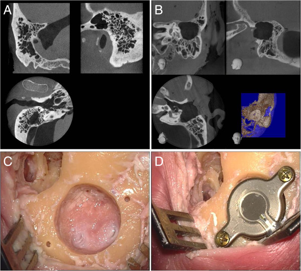
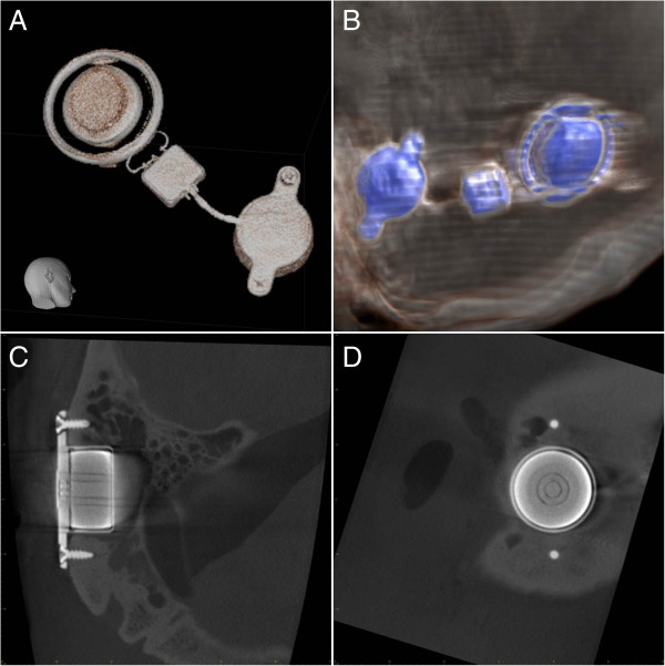
Methods: The Bonebridge was implanted unilaterally in two completely prepared human heads. The radiological imaging by means of CBCT, 64-slice CT, 1.5-T and 3.0-T MRI was conducted both preoperatively and postoperatively. The images were subsequently evaluated from both the ENT medical and nd radiological perspectives.
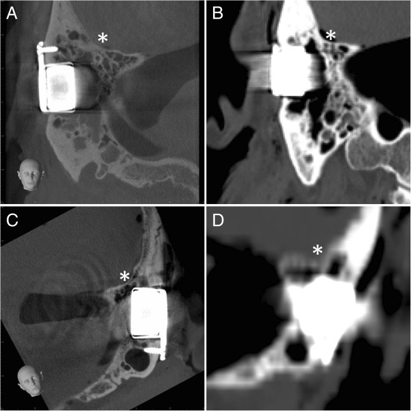
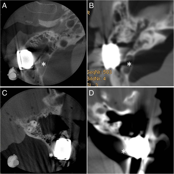
Results: As anticipated, no visualisation of the implant or of the petrosal bones could be realised on MRI because of the interactive technology and the magnet artefact. In contrast, an excellent evaluability of the implant itself as well as of the surrounding neurovascular structures (sinus sigmoideus, skull base, middle ear, inner ear, inner auditory canal) was exhibited in both the CT and in the CBCT.
Conclusion: The Bonebridge can be excellently imaged with the radiological imaging technologies of CT and CBCT. In the process, CBCT shows discrete advantages in comparison with CT. No relevant restrictions in image quality in the evaluation of the bony structures of the petrosal bones could be seen.
Figures
References
-
- Snik AF, Mylanus EA, Proops DW, Wolfaardt JF, Hodgetts WE, Somers T, Niparko JK, Wazen JJ, Sterkers O, Cremers CW, Tjellstrom A. Consensus statements on the BAHA system: where do we stand at present? Ann Otol Rhinol Laryngol Suppl. 2005;195:2–12. - PubMed
MeSH terms
LinkOut - more resources
Full Text Sources
Other Literature Sources
Medical
Research Materials
Miscellaneous
