. 2013 Oct;140(19):4041-50.
doi: 10.1242/dev.096255. Epub 2013 Sep 4.

Dynamic responses of endothelial cells to changes in blood flow during vascular remodeling of the mouse yolk sac

Affiliations

Dynamic responses of endothelial cells to changes in blood flow during vascular remodeling of the mouse yolk sac

Ryan S Udan et al. Development. 2013 Oct.

Abstract

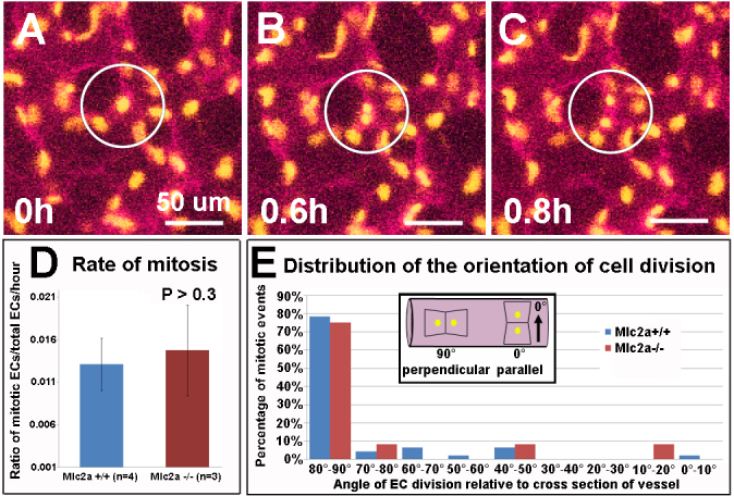
Despite extensive work showing the importance of blood flow in angiogenesis and vessel remodeling, very little is known about how changes in vessel diameter are orchestrated at the cellular level in response to mechanical forces. To define the cellular changes necessary for remodeling, we performed live confocal imaging of cultured mouse embryos during vessel remodeling. Our data revealed that vessel diameter increase occurs via two distinct processes that are dependent on normal blood flow: vessel fusions and directed endothelial cell migrations. Vessel fusions resulted in a rapid change in vessel diameter and were restricted to regions that experience the highest flow near the vitelline artery and vein. Directed cell migrations induced by blood flow resulted in the recruitment of endothelial cells to larger vessels from smaller capillaries and were observed in larger artery segments as they expanded. The dynamic and specific endothelial cell behaviors captured in this study reveal how sensitive endothelial cells are to changes in blood flow and how such responses drive vascular remodeling.

Keywords: Cell migration; Endothelial cell; Mouse; Vascular remodeling; Vessel fusion; Yolk sac.


Figures

Fig. 1.
Fig. 1.
Relationship between the number of endothelial cells and vessel diameter. (A-E) Flk1-myr::mCherry (magenta) and Flk1-H2B::eYFP (yellow) labeled vessels from flat-mounted E8.5 (A,B) and E9.5 (C,D) yolk sacs, with the distribution of vessel diameters at each stage (E). Proximal vessels (arrow) feed into distal vessels (arrowheads) (A-D). (F,G) The number of ECs per vessel diameter was measured by counting the number of YFP nuclei in a vessel segment. Vessel segment length [F, white bar (51.0 μm) or G, gray bar], diameter [F,G, green bar (81.96 μm)] and EC number were measured, and comparisons between EC number and diameter were made by adjusting the number of ECs per diameter vessel to a fixed vessel length (200 μm) (G). (H) The number of ECs per vessel diameter from E8.5, E9.5 and E10.5 yolk sacs (n=89 vessels from nine E8.5-10.5 mouse embryos). Trendlines represent best fits of the data. (I) EC density [number of ECs/area (mm2)] versus area (mm2) indicates no correlation between density and vessel segment area at any stage (R2 values are close to 0). (J) Box plots of density measurements evaluated at different stages show similar median values [line between lower quartile (light gray) and upper quartile (dark gray)] at E8.5 and E9.5, but the median is slightly decreased at E10.5. A significant difference in the variance (difference between the lower and upper extremes) was also detected between stages, as indicated in the text.
Fig. 2.
Fig. 2.
Quantification of the mitotic and apoptotic indices of ECs in normal flow and reduced flow yolk sac vasculature. The number of phospho-histone H3 (PH3)-positive cells or the number of cleaved caspase 3-positive cells that colabeled (arrows) with Flk1-H2B::eYFP were counted in normal flow (Mlc2a+/+) and reduced flow (Mlc2a-/-) mouse embryos at E8.5 (A-D or K-N) and E9.5 (F-I or P-S). The mitotic index (E,J) or apoptotic index (O,T) was determined at E8.5 and E9.5 by counting the total number of PH3-positive or cleaved caspase 3-positive ECs among total ECs. Error bars represent s.d. For the mitotic index, n=3 yolk sacs per stage and per genotype, with a total of 20 fields of view per yolk sac. For the apoptotic index, n=3 yolk sacs per stage and per genotype, with a total of eight fields of view per yolk sac.
Fig. 3.
Fig. 3.
Live imaging to quantify the orientation of cell divisions and mitotic indices in Mlc2a+/+ versus Mlc2a-/- vessels. (A-C) Time-lapse movies of Flk1-myr::mCherry (magenta) and Flk1-H2B::eYFP (yellow) labeled vessels capture mitotic events that take place over 0-0.8 hours (separation of YFP-labeled nuclei, circled). (D) The mitotic index (number of mitotic events/average number of ECs per movie)/(time of EC in mitosis/total time of movie) was determined from Mlc2a+/+ and Mlc2a-/- movies, and no statistically significant difference in the rate of mitosis was observed (P>0.3, t-test). Error bars indicate s.d. (E) The distribution of the angles of EC division, relative to the cross-section of the vessel (0°, arrow in inset), was plotted in Mlc2a-/- and Mlc2a+/+ vessels from time-lapse movies.
Fig. 4.
Fig. 4.
Time-lapse analysis of changes in vessel diameter. Time-lapse movie sequences (starting at E8.5) of Flk1-myr::mCherry; Flk1-H2B::eYFP labeled ECs from Mlc2a+/+ proximal arteries (A-D), Mlc2a-/- proximal arteries (F-I), Mlc2a+/+ distal capillaries (K-N) and Mlc2a+/+ proximal veins (P-S). Detail of the boxed regions in A, F, K and P are shown for select time-lapse sequences. Changes in vessel diameters (white bar) are displayed graphically over time (E,J,O,T). Rapid changes in vessel diameters (red arrows) were coincident with the fusion of two neighboring vessels and the loss of an avascular space between them, as in the Mlc2a+/+ proximal artery (A-D) or the Mlc2a+/+ proximal vein (P-S), but such rapid changes were not seen in Mlc2a-/- vessels (F-I) or Mlc2a+/+ distal capillaries (K-N). A slower rate of change was also observed during Mlc2a+/+ proximal artery remodeling, as there was a steady rate of diameter increase in addition to the rapid change associated with fusions (A-D). This was not observed in other groups that either exhibited reduced vessel growth (Mlc2a-/- vessels, F-I), no net change in diameter (Mlc2a+/+ distal capillaries, K-N) or where the only diameter increase that occurred correlated with the fusion event (Mlc2a+/+ proximal vein, P-S).
Fig. 5.
Fig. 5.
Blood flow profiles at different stages of remodeling from developing arterial, venous and capillary vessels. Fast line-scan confocal imaging was used to track ε-Globin-GFP labeled erythrocytes to measure blood velocities in developing arterial and venous vessels. Blood velocities were plotted over time (∼1.4 seconds) in developing arteries, capillaries and veins of late E8.25 (A-C), E8.75 (D-F) and E9.5 (G-I) mouse embryos.
Fig. 6.
Fig. 6.
ECs show regional differences in migration. Images from time-lapse sequences from 0, 4 or 6 hours after the start of the sequence, showing the continuous migration tracks (white lines) of selected ECs. The direction of blood flow is indicated by green arrows. Blue circles indicate the position of the tracked EC at the time of the frame of the time-lapse sequence, and the red ‘X’ indicates the end of each migration track. Tracks of ECs within the developing arteries of wild-type embryos showed migration from low-flow interconnected capillaries towards and into the growing arteries with high flow (A-D), and showed proximal migration within high-flow arteries (E-H). By contrast, migration tracks of ECs in the proximal arteries of Mlc2a-/- embryos (I-L), in wild-type capillaries (M-P) and in wild-type proximal veins (Q-T) did not appear to have a consistent direction or extend very far from the start position.
Fig. 7.
Fig. 7.
ECs exhibit increased motility and directional migration in interconnected vessels adjacent to and within vessels of high flow. (A-D) EC tracks (colored lines) and displacement vectors (gray arrows) in selected arterial or capillary regions are shown for: (A) low-flow vessels (LFV) from Mlc2a-/- embryos (group I); (B) LFV from Mlc2a+/+ embryos (group II); (C) low-flow vessels interconnected to high-flow vessels (LFV-HFV) from Mlc2a+/+ embryos (group III); and (D) high-flow vessels (HFV) from Mlc2a+/+ embryos (group IV). (E-H) Average total displacement (E), average total track length (F) and average tortuosity (G) of EC migration tracks were measured for each group. Significant differences in the average displacement and average tortuosity were seen for groups I/II compared with groups III/IV. Differences in the angles of EC displacement tracks, relative to the region of higher flow, are shown for groups III and IV in H. A non-linear regression was performed to demonstrate a Gaussian distribution for both groups. Error bars indicate s.d.
Fig. 8.
Fig. 8.
Model of yolk sac vascular remodeling in response to changes in blood flow. (A) Blood enters the vascular plexus at E8.5 through the vitelline artery. In the proximal arterial region of the yolk sac, a randomly chosen, preferred path of flow forms. Blood is then diverted to the distal capillaries and is ultimately collected by the vitelline vein. Vessel remodeling occurs by the redistribution of ECs via vessel fusions (circles) and directed EC migrations (white arrows in B,C), resulting in a rapid and a slower change in vessel diameter, respectively. Proximal vessels that are exposed to high peak blood velocities and high resistances undergo vessel fusion (circles) with neighboring vessels. (B,C) By E9.0-9.5, fusions help to establish vessel hierarchy, resulting in proximal arteries with pulsatile/high velocity flows, distal capillaries with pulsatile/low velocity flows, and proximal veins with steadier/low velocity flows. The amount of force that is exhibited by these flow profiles correlates well with observed directed migrations that occur from low-flow vessels to high-flow/growing arteries, and upstream of flow in high-flow/growing arteries - events that are absent in distal capillaries and proximal veins. The unique responses that ECs exhibit in different regions might explain not only how vessel hierarchies form, but also how arterial and venous morphologies are established.

References

    1. Argraves W. S., Drake C. J. (2005). Genes critical to vasculogenesis as defined by systematic analysis of vascular defects in knockout mice. Anat. Rec. A Discov. Mol. Cell. Evol. Biol. 286, 875–884 - PubMed
    1. Armstrong J. J., Larina I. V., Dickinson M. E., Zimmer W. E., Hirschi K. K. (2010). Characterization of bacterial artificial chromosome transgenic mice expressing mCherry fluorescent protein substituted for the murine smooth muscle alpha-actin gene. Genesis 48, 457–463 - PMC - PubMed
    1. Califano J. P., Reinhart-King C. A. (2010). Exogenous and endogenous force regulation of endothelial cell behavior. J. Biomech. 43, 79–86 - PubMed
    1. Chatzizisis Y. S., Coskun A. U., Jonas M., Edelman E. R., Feldman C. L., Stone P. H. (2007). Role of endothelial shear stress in the natural history of coronary atherosclerosis and vascular remodeling: molecular, cellular, and vascular behavior. J. Am. Coll. Cardiol. 49, 2379–2393 - PubMed
    1. Chiu J. J., Chien S. (2011). Effects of disturbed flow on vascular endothelium: pathophysiological basis and clinical perspectives. Physiol. Rev. 91, 327–387 - PMC - PubMed

Publication types

MeSH terms

Substances

LinkOut - more resources