

The potential role of modern US in the follow-up of patients with retroperitoneal fibrosis
- PMID: 24004976
- PMCID: PMC4463242
- DOI: 10.5152/dir.2013.13132
The potential role of modern US in the follow-up of patients with retroperitoneal fibrosis
Abstract
Purpose: We aimed to evaluate a standardized ultrasonography (US) algorithm for the visualization of pathologic para-aortic tissue in retroperitoneal fibrosis (RPF).
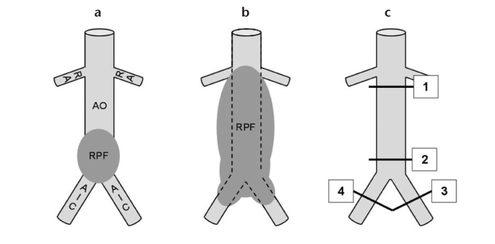
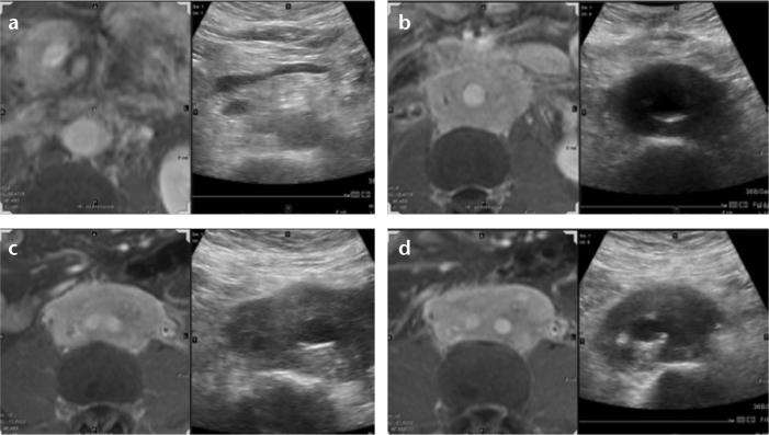
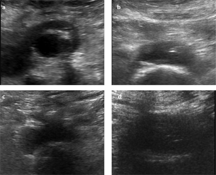
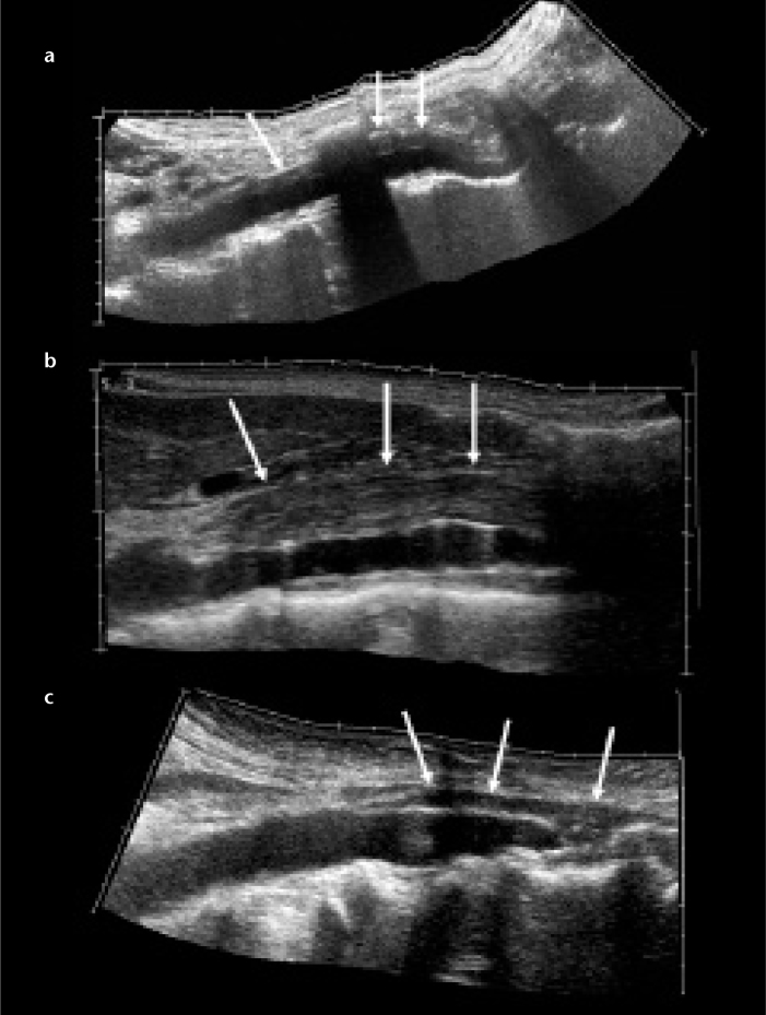
Materials and methods: Thirty-five patients with lumbar RPF of typical extent, as determined by abdominal magnetic resonance imaging, were included. Examinations were conducted using standardized abdominal US with axial sections obtained at the levels of the renal arteries, aortic bifurcation, and both common iliac arteries. Imaging of each section was acquired with fundamental B-mode (US) and tissue harmonic imaging, respectively. In addition, we examined RPF visualized using extended field-of-view US.
Results: Tissue harmonic imaging adequately visualized RPF of typical extent in 33 patients (94.2%). Excellent and good visualization with mild artifacts were achieved in 25 (71.4%) and six (17.1%) patients, respectively. When RPF spread along the iliac arteries, excellent visualization was achieved in 38.7% for the left side and 34.5% for the right side. There were significantly fewer diagnostic examinations for the right iliac (27.6%) than for the left one (9.7%) (P = 0.016). Overall, harmonic imaging achieved significantly better visualization than fundamental B-Mode (P < 0.001).
Conclusion: We described the first systematic evaluation of RPF visualization by modern US techniques. The best imaging quality was found in the typical RPF location, at the level of the aortic bifurcation. These results advocate for the presented US algorithm as an efficient follow-up alternative to cross-sectional imaging in RPF patients.
Figures
Similar articles
-
Imaging appearance of fibrosing diseases of the retroperitoneum: can a definitive diagnosis be made?Abdom Radiol (NY). 2018 May;43(5):1204-1214. doi: 10.1007/s00261-017-1282-5. Abdom Radiol (NY). 2018. PMID: 28849414
-
Retroperitoneal fibrosis: role of imaging in diagnosis and follow-up.Radiographics. 2013 Mar-Apr;33(2):535-52. doi: 10.1148/rg.332125085. Radiographics. 2013. PMID: 23479712
-
Differentiation of Lymphoma Presenting as Retroperitoneal Mass and Retroperitoneal Fibrosis: Evaluation with Multidetector-row Computed Tomography.Chin Med J (Engl). 2017 Mar 20;130(6):691-697. doi: 10.4103/0366-6999.201606. Chin Med J (Engl). 2017. PMID: 28303852 Free PMC article.
-
[Clinical characteristics of IgG4 related retroperitoneal fibrosis: a report of 5 cases and literature review].Beijing Da Xue Xue Bao Yi Xue Ban. 2015 Aug 18;47(4):622-7. Beijing Da Xue Xue Bao Yi Xue Ban. 2015. PMID: 26284398 Review. Chinese.
-
Idiopathic retroperitoneal fibrosis (RPF): clinical features of 61 cases and literature review.Clin Rheumatol. 2011 May;30(5):601-5. doi: 10.1007/s10067-010-1580-6. Epub 2010 Oct 19. Clin Rheumatol. 2011. PMID: 20957401 Review.
Cited by
-
[Diseases of connective tissue in IgG4-associated autoimmune diseases].Radiologe. 2016 Dec;56(12):1061-1068. doi: 10.1007/s00117-016-0183-x. Radiologe. 2016. PMID: 27882402 Review. German.
-
[Diagnosis and treatment of retroperitoneal fibrosis].Urologe A. 2016 Jun;55(6):732-40. doi: 10.1007/s00120-016-0081-x. Urologe A. 2016. PMID: 27168039 Review. German.
-
[New (and old) aspects of retroperitoneal fibrosis].Urologe A. 2017 Jul;56(7):887-894. doi: 10.1007/s00120-017-0428-y. Urologe A. 2017. PMID: 28643106 Review. German.
References
-
- Burkhardt Soares S, Fehr A, Brandt AS, et al. Retroperitonele fibrose. Aktuelle Urologie. 2007;38:221–231. - PubMed
-
- Cronin CG, Lohan DG, Blake MA, Roche C, McCarthy P, Murphy JM. Retroperitoneal fibrosis: a review of clinical features and imaging findings. Am J Roentgenol. 2008;191:423–431. - PubMed
-
- Vaglio A, Salvarani C, Buzio C. Retroperitoneal fibrosis. Lancet. 2006;367:241–251. - PubMed
-
- Amis ES., Jr Retroperitoneal fibrosis. Am J Roentgenol. 1991;157:321–329. - PubMed
-
- Vivas I, Nicolás AI, Velázquez P, Elduayen B, Fernández-Villa T, Martínez-Cuesta A. Retroperitoneal fibrosis: typical and atypical manifestations. Br J Radiol. 2000;73:214–222. - PubMed
MeSH terms
LinkOut - more resources
Full Text Sources
Other Literature Sources
