

Peripheral cardiac sympathetic hyperactivity in cardiovascular disease: role of neuropeptides
- PMID: 24005254
- PMCID: PMC3882692
- DOI: 10.1152/ajpregu.00118.2013
Peripheral cardiac sympathetic hyperactivity in cardiovascular disease: role of neuropeptides
Abstract
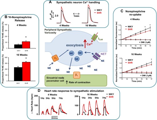
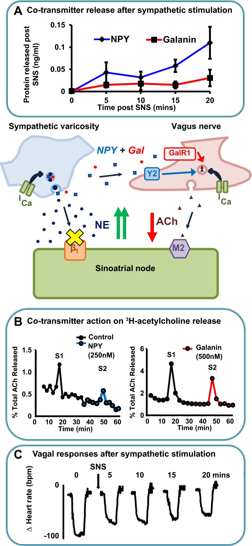
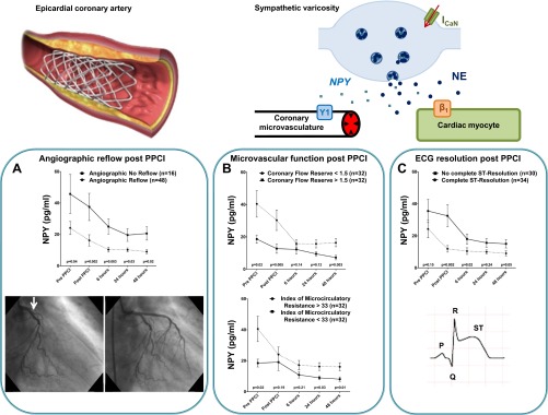
High levels of sympathetic drive in several cardiovascular diseases including postmyocardial infarction, chronic congestive heart failure and hypertension are reinforced through dysregulation of afferent input and central integration of autonomic balance. However, recent evidence suggests that a significant component of sympathetic hyperactivity may also reside peripherally at the level of the postganglionic neuron. This has been studied in depth using the spontaneously hypertensive rat, an animal model of genetic essential hypertension, where larger neuronal calcium transients, increased release and impaired reuptake of norepinephrine in neurons of the stellate ganglia lead to a significant tachycardia even before hypertension has developed. The release of additional sympathetic cotransmitters during high levels of sympathetic drive can also have deleterious consequences for peripheral cardiac parasympathetic neurotransmission even in the presence of β-adrenergic blockade. Stimulation of the cardiac vagus reduces heart rate, lowers myocardial oxygen demand, improves coronary blood flow, and independently raises ventricular fibrillation threshold. Recent data demonstrates a direct action of the sympathetic cotransmitters neuropeptide Y (NPY) and galanin on the ability of the vagus to release acetylcholine and control heart rate. Moreover, there is as a strong correlation between plasma NPY levels and coronary microvascular function in patients with ST-elevation myocardial infarction being treated with primary percutaneous coronary intervention. Antagonists of the NPY receptors Y1 and Y2 may be therapeutically beneficial both acutely during myocardial infarction and also during chronic heart failure and hypertension. Such medications would be expected to act synergistically with β-blockers and implantable vagus nerve stimulators to improve patient outcome.
Keywords: autonomic nervous system; cardiac; hypertension; myocardial infarction; neuropeptide Y.
Figures
Similar articles
-
Cardiac sympathetic dysfunction in the prehypertensive spontaneously hypertensive rat.Am J Physiol Heart Circ Physiol. 2013 Oct 1;305(7):H980-6. doi: 10.1152/ajpheart.00255.2013. Epub 2013 Aug 2. Am J Physiol Heart Circ Physiol. 2013. PMID: 23913706 Free PMC article.
-
The cardiac sympathetic co-transmitter neuropeptide Y is pro-arrhythmic following ST-elevation myocardial infarction despite beta-blockade.Eur Heart J. 2020 Jun 14;41(23):2168-2179. doi: 10.1093/eurheartj/ehz852. Eur Heart J. 2020. PMID: 31834357 Free PMC article.
-
Circulating noradrenaline leads to release of neuropeptide Y from cardiac sympathetic nerve terminals via activation of β-adrenergic receptors.J Physiol. 2025 Mar;603(7):1911-1921. doi: 10.1113/JP285945. Epub 2024 Feb 14. J Physiol. 2025. PMID: 38352977
-
Autonomic control of the heart: going beyond the classical neurotransmitters.Exp Physiol. 2015 Apr 1;100(4):354-8. doi: 10.1113/expphysiol.2014.080184. Epub 2014 Nov 20. Exp Physiol. 2015. PMID: 25344273 Free PMC article. Review.
-
Neuromodulators of peripheral cardiac sympatho-vagal balance.Exp Physiol. 2009 Jan;94(1):46-53. doi: 10.1113/expphysiol.2008.044776. Epub 2008 Oct 22. Exp Physiol. 2009. PMID: 18945757 Review.
Cited by
-
Deletion of Neuropeptide Y Attenuates Cardiac Dysfunction and Apoptosis During Acute Myocardial Infarction.Front Pharmacol. 2019 Oct 24;10:1268. doi: 10.3389/fphar.2019.01268. eCollection 2019. Front Pharmacol. 2019. PMID: 31708788 Free PMC article.
-
The Effects of Neuropeptide Y Overexpression on the Mouse Model of Doxorubicin-Induced Cardiotoxicity.Cardiovasc Toxicol. 2020 Jun;20(3):328-338. doi: 10.1007/s12012-019-09557-2. Cardiovasc Toxicol. 2020. PMID: 31811615 Free PMC article.
-
The hydrophobic C-terminal sequence of transthyretin affects its catalytic kinetics towards amidated neuropeptide Y.FEBS Open Bio. 2019 Mar 4;9(4):594-604. doi: 10.1002/2211-5463.12604. eCollection 2019 Apr. FEBS Open Bio. 2019. PMID: 30984535 Free PMC article.
-
Ultrastructural Correlates of Enhanced Norepinephrine and Neuropeptide Y Cotransmission in the Spontaneously Hypertensive Rat Brain.ASN Neuro. 2015 Oct 29;7(5):1759091415610115. doi: 10.1177/1759091415610115. Print 2015 Sep-Oct. ASN Neuro. 2015. PMID: 26514659 Free PMC article.
-
Cardiovascular Alterations and Management of Patients With White Coat Hypertension: A Meta-Analysis.Front Pharmacol. 2020 Sep 17;11:570101. doi: 10.3389/fphar.2020.570101. eCollection 2020. Front Pharmacol. 2020. PMID: 33041810 Free PMC article.
References
-
- Adrian TE, Gu J, Allen JM, Tatemoto K, Polak JM, Bloom SR. Neuropeptide Y in the human male genital tract. Life Sci 35: 2643–2648, 1984 - PubMed
-
- Anderson EA, Sinkey CA, Lawton WJ, Mark AL. Elevated sympathetic nerve activity in borderline hypertensive humans: evidence from direct intraneural recordings. Hypertension 14: 177–183, 1989 - PubMed
-
- Armour JA. Potential clinical relevance of the “little brain” on the mammalian heart. Exp Physiol 93: 165–176, 2008 - PubMed
-
- Boudaka A, Worl J, Shiina T, Shimizu Y, Takewaki T, Neuhuber WL. Galanin modulates vagally induced contractions in the mouse oesophagus. Neurogastroenterol Motil 21: 180–188, 2009 - PubMed
-
- Bowers CW, Zigmond RE. Localization of neurons in the rat superior cervical ganglion that project into different postganglionic trunks. J Comp Neurol 185: 381–391, 1979 - PubMed
Publication types
MeSH terms
Substances
Grants and funding
LinkOut - more resources
Full Text Sources
Other Literature Sources
Research Materials
Miscellaneous