. 2013 Sep 4;33(36):14392-405.
doi: 10.1523/JNEUROSCI.2045-13.2013.

A reorganized GABAergic circuit in a model of epilepsy: evidence from optogenetic labeling and stimulation of somatostatin interneurons

Affiliations

A reorganized GABAergic circuit in a model of epilepsy: evidence from optogenetic labeling and stimulation of somatostatin interneurons

Zechun Peng et al. J Neurosci. .

Abstract

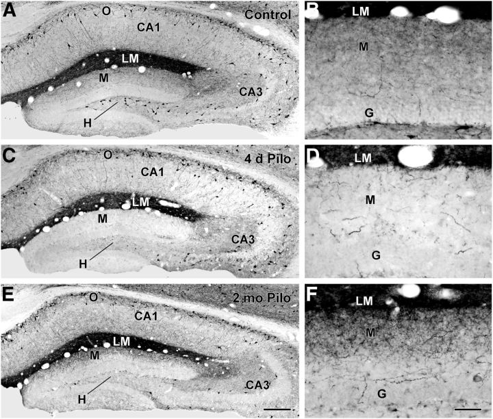
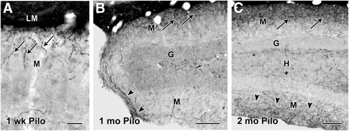
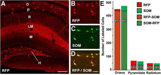
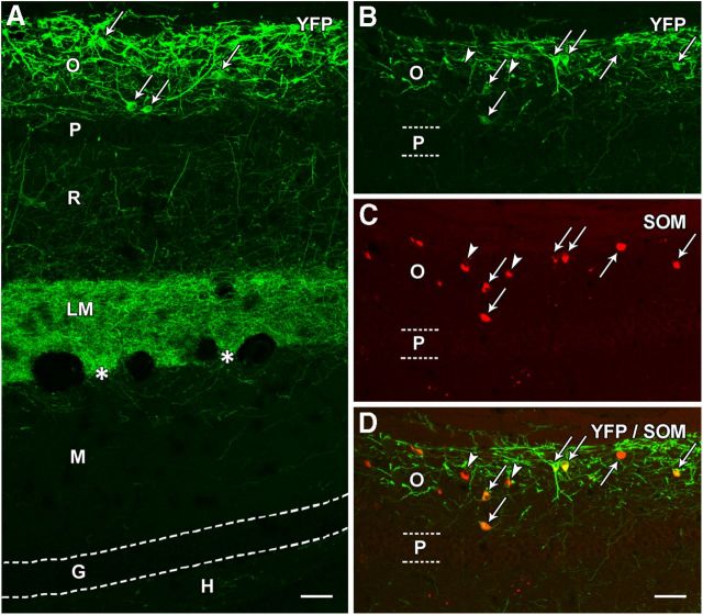
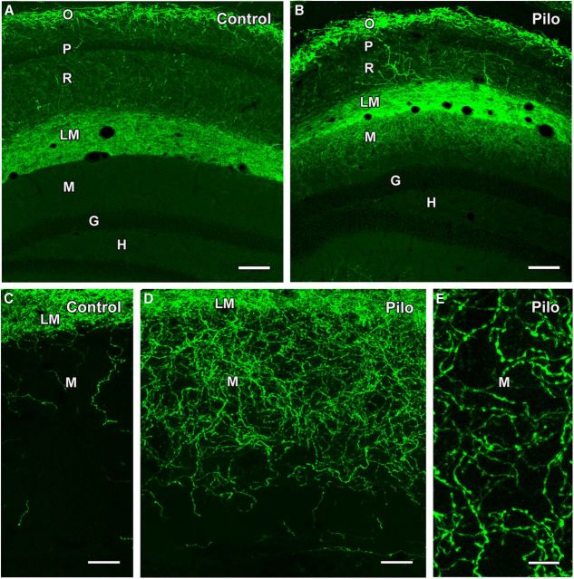
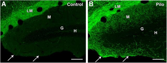
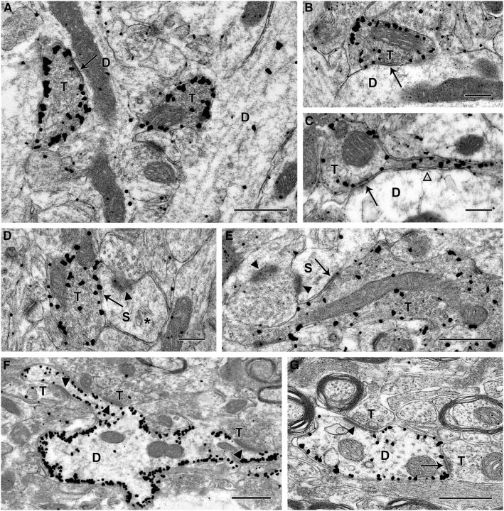
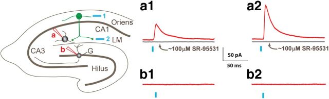
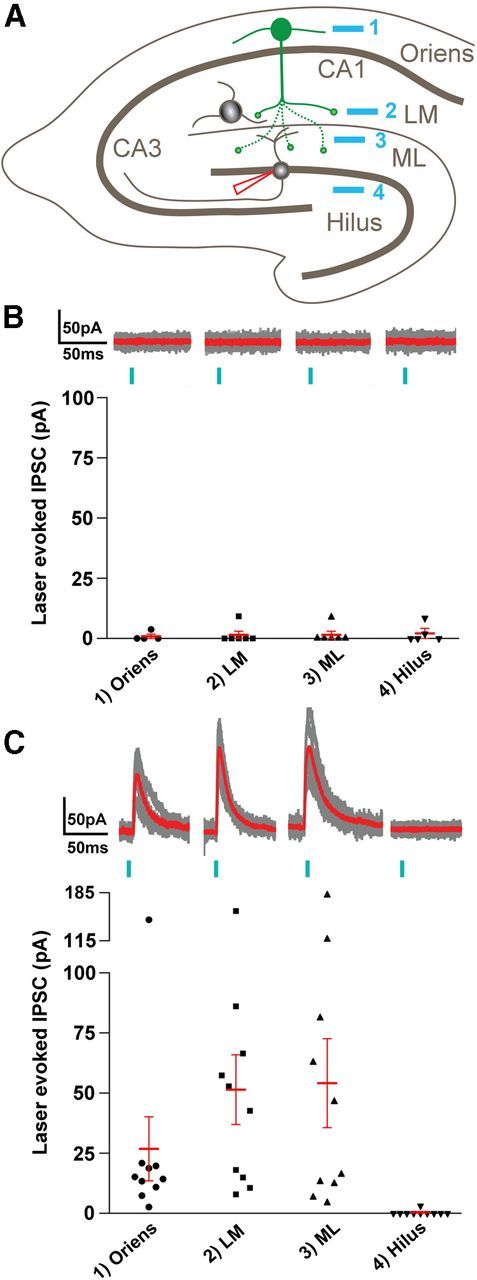
Axonal sprouting of excitatory neurons is frequently observed in temporal lobe epilepsy, but the extent to which inhibitory interneurons undergo similar axonal reorganization remains unclear. The goal of this study was to determine whether somatostatin (SOM)-expressing neurons in stratum (s.) oriens of the hippocampus exhibit axonal sprouting beyond their normal territory and innervate granule cells of the dentate gyrus in a pilocarpine model of epilepsy. To obtain selective labeling of SOM-expressing neurons in s. oriens, a Cre recombinase-dependent construct for channelrhodopsin2 fused to enhanced yellow fluorescent protein (ChR2-eYFP) was virally delivered to this region in SOM-Cre mice. In control mice, labeled axons were restricted primarily to s. lacunosum-moleculare. However, in pilocarpine-treated animals, a rich plexus of ChR2-eYFP-labeled fibers and boutons extended into the dentate molecular layer. Electron microscopy with immunogold labeling demonstrated labeled axon terminals that formed symmetric synapses on dendritic profiles in this region, consistent with innervation of granule cells. Patterned illumination of ChR2-labeled fibers in s. lacunosum-moleculare of CA1 and the dentate molecular layer elicited GABAergic inhibitory responses in dentate granule cells in pilocarpine-treated mice but not in controls. Similar optical stimulation in the dentate hilus evoked no significant responses in granule cells of either group of mice. These findings indicate that under pathological conditions, SOM/GABAergic neurons can undergo substantial axonal reorganization beyond their normal territory and establish aberrant synaptic connections. Such reorganized circuitry could contribute to functional deficits in inhibition in epilepsy, despite the presence of numerous GABAergic terminals in the region.


Figures

Figure 1.
Figure 1.
SOM immunoreactivity in control (A–B) and pilocarpine (Pilo)-treated (C–F) mice. A, B, In a control mouse, cell bodies of SOM neurons are concentrated in s. oriens (O) of CA1 and CA3 of the hippocampus and the hilus (H) of the dentate gyrus. A high concentration of SOM fibers and terminals creates a dark band of labeling in s. lacunosum-moleculare (LM) of CA1. Lower concentrations of fibers and terminals are evident in the outer molecular layer (M) of the dentate gyrus. C, D, At 4 d following pilocarpine-induced status epilepticus, SOM neurons in the dentate hilus are severely depleted. A marked loss of SOM-labeled fibers and terminals is also evident in the molecular layer, consistent with the normal origin of these fibers from SOM neurons in the hilus. Many SOM neurons remain in s. oriens of CA1, and the band of dense labeling remains in s. lacunosum-moleculare. E, F, At 2 months following pilocarpine-induced seizures, cell bodies of SOM neurons in the hilus remain depleted. However, SOM-labeled fibers and terminals are evident in the outer molecular layer of the dentate gyrus. Scale bars: (in E) A,C,E, 200 μm; (in F) B,D,F, 50 μm. G, granule cell layer.
Figure 2.
Figure 2.
Patterns of SOM-immunoreactive fibers at different intervals following pilocarpine treatment. A, As early as 1 week after status epilepticus, a small number of SOM-labeled fibers (arrows), often with a vertical orientation, are evident in the outer part of the molecular layer (M), adjacent to s. lacunosum-moleculare (LM). B, At 1 month following pilocarpine-induced seizures, increased SOM labeling is evident in the outer molecular layer (arrows), and some fibers (arrowheads) appear to be extending in a dorsal to ventral direction around the crest of the dentate gyrus. C, At 2 months following pilocarpine-induced seizures, increased SOM labeling is evident in the molecular layer of the suprapyramidal blade of the dentate gyrus (arrows), and labeling is also evident in the molecular layer of the infrapyramidal blade (arrowheads). Labeled cell bodies remain depleted in the hilus (H). Scale bars: A, 25 μm; B, C, 100 μm. G, granule cell layer.
Figure 3.
Figure 3.
Localization of endogenous RFP and SOM immunoreactivity in SOM-Cre-Ai9 mice. A, RFP, a reporter of Cre expression in these mice, is present in the cell bodies of numerous neurons in s. oriens (O) in CA1 and the dentate hilus (H), with relatively few labeled neurons within s. pyramidale (P) and s. radiatum (R). A dense band of labeled fibers and terminals is evident in s. lacunosum-moleculare (LM), and more moderate labeling of fibers is present in the outer molecular layer (M) of the dentate gyrus. This labeling closely resembles the normal patterns of SOM immunoreactivity (Fig. 1A). B–D, In sections processed for SOM immunohistochemistry, the majority of RFP-labeled neurons in s. oriens are also immunolabeled for SOM, as demonstrated by the merged images. All cell bodies in this region of s. oriens are double labeled. E, Quantitative analysis of single- and double-labeled cells in CA1 demonstrated a high correspondence between RFP and SOM labeling in s. oriens, where the majority of RFP-labeled cells were located. Lower percentages of double-labeled neurons were found in s. pyramidale and s. radiatum, but the number of neurons in these regions was substantially smaller than in s. oriens. Scale bars: A, 200 μm; B–D, 50 μm.
Figure 4.
Figure 4.
Localization of eYFP in a normal SOM-Cre mouse following viral delivery of Cre-dependent ChR2-eYFP construct to s. oriens. A, At 3 weeks after injection, eYFP labeling is prominent in neuronal cell bodies (examples at arrows) and processes in s. oriens (O) of CA1. A few labeled processes extend through the adjacent pyramidal cell layer (P) and traverse s. radiatum (R). A dense plexus of fibers is present throughout s. lacunosum-moleculare (LM) but ends sharply at the hippocampal fissure (asterisks). Very limited fiber labeling is evident in the adjacent molecular layer (M) of the dentate gyrus. Virtually no labeling is present in the granule cell layer (G) or hilus (H) of the dentate gyrus, indicating that the viral vector did not reach the hilar region where additional Cre-expressing neurons are located in normal mice. B–D, In sections immunolabeled for SOM, essentially all eYFP-expressing neurons were SOM neurons (arrows), as demonstrated by the merged images. However, a few SOM neurons in the region did not appear to be labeled for eYFP (arrowheads). Scale bars: 50 μm.
Figure 5.
Figure 5.
Comparison of ChR2-eYFP labeling in control and pilocarpine (Pilo)-treated mice following viral vector injection in s. oriens of SOM-Cre mice. A, B, In a control mouse, numerous neurons in s. oriens (O) of CA1 are labeled for eYFP, with very limited cell body labeling in s. pyramidale (P) and s. radiatum (R). A dense plexus of labeled fibers is evident in s. lacunosum-moleculare (LM) but does not extend into the adjacent molecular layer (M) of the dentate gyrus. No labeling is evident in the granule cell layer (G) or hilus (H). In a pilocarpine-treated mouse, similar eYFP labeling is evident in s. oriens and s. lacunosum moleculare, but, in contrast to the control, substantial labeling extends into the outer molecular layer of the dentate gyrus. C, D, At higher magnification, limited numbers of eYFP-labeled fibers are present in the outer molecular layer in the control mouse. In contrast, a rich plexus of labeled fibers is evident throughout the outer two-thirds of the molecular layer in a pilocarpine-treated mouse. E, Labeled fibers in the molecular layer of a pilocarpine-treated mouse exhibit numerous small swellings separated by thin labeled segments, suggesting axons with en passant terminals. Scale bars: A, B, 100 μm; C, D, 25 μm; E, 10 μm.
Figure 6.
Figure 6.
Comparison of ChR2-eYFP labeling in control and pilocarpine (Pilo)-treated mice in both blades of the dentate gyrus. A, In a control mouse, only a few labeled fibers are evident in the outer molecular layer (M) of the suprapyramidal (inner) blade of the dentate gyrus, adjacent to s. lacunosum-moleculare (LM), and even fewer labeled fibers are present in the infrapyramidal (outer) blade (arrows). No labeling of cell bodies is evident in the hilus (H) or granule cell layer (G). B, In a pilocarpine-treated mouse, a rich plexus of eYFP-labeled fibers extends through much of the molecular layer of the inner blade of the dentate gyrus, and the fibers appear to continue around the crest and into the molecular layer of the outer blade (arrows). As in the control, there is a lack of cell body labeling in the hilus and granule cell layer. Scale bars: 100 μm.
Figure 7.
Figure 7.
Electron micrographs of immunogold labeling of eYFP at 3 weeks following viral-mediated transfection of ChR2-eYFP in s. oriens of CA1 in a pilocarpine-treated SOM-Cre mouse. A–C, Immunogold-labeled terminals (T) in the dentate molecular layer form distinct symmetric synaptic contacts (arrows) with medium to small dendritic shafts (D). Immunogold particles are frequently concentrated near the periphery of the labeled axon terminals. The labeled terminals contain mitochondrial profiles and numerous synaptic vesicles, consistent with functional terminals. C, A labeled axon terminal is in continuity with the thin preterminal segment of its axon (open arrowhead). D, E, eYFP-labeled terminals also form symmetric synaptic contacts (arrows) with dendritic spines (S) in the dentate molecular layer. These spines form asymmetric synaptic contacts (arrowheads) with unlabeled terminals. In D, the spine contains a spine apparatus (*), and in E, the labeled terminal is relatively large and forms an en passant synapse with the spine (S). F, G, In s. oriens of CA1, eYFP-labeled dendrites appear outlined by immunogold particles, which are located near the inner face of the plasma membrane. These dendrites commonly received synaptic contacts. F, Unlabeled terminals (T) form asymmetric synapses (arrowheads) with a labeled dendrite, and two of these contacts are with an elongated spine-like process that extends from the larger dendritic shaft. G, A labeled dendrite receives both an asymmetric (arrowhead) and a probable symmetric (arrow) synaptic contact from unlabeled axon terminals. Scale bars: A, E, F, G, 0.5 μm; B–D, 0.25 μm.
Figure 8.
Figure 8.
Regional stimulation of ChR2 with patterned illumination in control mice demonstrates that neurons in s. oriens contact interneurons in s. lacunosum-moleculare (LM) but not granule cells (G) in the dentate gyrus. On the left is a schematic illustrating a hippocampal slice and the locations of ChR2-expressing neurons in s. oriens (green) and the stimulation regions targeted by blue laser light (blue numbered boxes). Whole-cell recordings from either LM interneurons (red pipette, site a) or dentate gyrus granule cells (red pipette, site b) were made in control slices. To the right are displayed laser-evoked IPSCs (red traces) recorded in response to stimulation at the two positions (1, oriens; 2, LM) in an LM interneuron (a1 and a2) and in a dentate gyrus granule cell (b1 and b2). Following application of ∼100 μm SR-95531, the postsynaptic response to laser pulses was abolished (gray traces in a1 and a2).
Figure 9.
Figure 9.
Reorganization of GABAergic circuitry in pilocarpine-treated mice. A, Schematic illustrating the patterned illumination laser stimuli in s. oriens (site 1), s. lacunosum-moleculare of CA1 (LM, site 2), dentate molecular layer (ML, site 3), and hilus (site 4), respectively, as well as whole-cell recordings used in granule cells (red pipette). B, Summary of stimulating these different locations in control mice (n = 4–6 cells). Top, Each part shows individual IPSCs (gray) superimposed on an average response (red) for the indicated locations (1–4). Bottom, A scatter plot showing the mean amplitudes of laser-evoked IPSCs for the indicated locations. C, Summary of responses in pilocarpine-treated mice (n = 6–10 cells). Top, Individual (gray) and average (red) responses for each of the sites (1–4). Bottom, Summarizes the mean IPSC amplitudes for each cell with the mean ± SEM. ANOVA analysis with post hoc Nemenyi test showed significant differences (p < 0.05) within the pilocarpine groups between all locations and the hilus as well as between normal and pilocarpine groups for the LM (p < 0.025) and ML (p < 0.05) locations.

Similar articles

Cited by

References

    1. Babb TL, Kupfer WR, Pretorius JK, Crandall PH, Levesque MF. Synaptic reorganization by mossy fibers in human epileptic fascia dentata. Neuroscience. 1991;42:351–363. doi: 10.1016/0306-4522(91)90380-7. - DOI - PubMed
    1. Bausch SB. Axonal sprouting of GABAergic interneurons in temporal lobe epilepsy. Epilepsy Behav. 2005;7:390–400. doi: 10.1016/j.yebeh.2005.07.019. - DOI - PubMed
    1. Blasco-Ibáñez JM, Freund TF. Synaptic input of horizontal interneurons in stratum oriens of the hippocampal CA1 subfield: structural basis of feed-back activation. Eur J Neurosci. 1995;7:2170–2180. doi: 10.1111/j.1460-9568.1995.tb00638.x. - DOI - PubMed
    1. Boulland JL, Ferhat L, Tallak Solbu T, Ferrand N, Chaudhry FA, Storm-Mathisen J, Esclapez M. Changes in vesicular transporters for gamma-aminobutyric acid and glutamate reveal vulnerability and reorganization of hippocampal neurons following pilocarpine-induced seizures. J Comp Neurol. 2007;503:466–485. doi: 10.1002/cne.21384. - DOI - PubMed
    1. Buckmaster PS, Dudek FE. Neuron loss, granule cell axon reorganization, and functional changes in the dentate gyrus of epileptic kainate-treated rats. J Comp Neurol. 1997;385:385–404. doi: 10.1002/(SICI)1096-9861(19970901)385:3<385::AID-CNE4>3.3.CO%3B2-Y. - DOI - PubMed

Publication types

MeSH terms