. 2013 Sep 4;33(36):14526-33.
doi: 10.1523/JNEUROSCI.0631-13.2013.

Simultaneous EEG and fMRI reveals a causally connected subcortical-cortical network during reward anticipation

Affiliations

Simultaneous EEG and fMRI reveals a causally connected subcortical-cortical network during reward anticipation

Michael M Plichta et al. J Neurosci. .

Abstract

Electroencephalography (EEG) and functional magnetic resonance imaging (fMRI) have been used to study the neural correlates of reward anticipation, but the interrelation of EEG and fMRI measures remains unknown. The goal of the present study was to investigate this relationship in response to a well established reward anticipation paradigm using simultaneous EEG-fMRI recording in healthy human subjects. Analysis of causal interactions between the thalamus (THAL), ventral-striatum (VS), and supplementary motor area (SMA), using both mediator analysis and dynamic causal modeling, revealed that (1) THAL fMRI blood oxygenation level-dependent (BOLD) activity is mediating intermodal correlations between the EEG contingent negative variation (CNV) signal and the fMRI BOLD signal in SMA and VS, (2) the underlying causal connectivity network consists of top-down regulation from SMA to VS and SMA to THAL along with an excitatory information flow through a THAL→VS→SMA route during reward anticipation, and (3) the EEG CNV signal is best predicted by a combination of THAL fMRI BOLD response and strength of top-down regulation from SMA to VS and SMA to THAL. Collectively, these findings represent a likely neurobiological mechanism mapping a primarily subcortical process, i.e., reward anticipation, onto a cortical signature.


Figures

Figure 1.
Figure 1.
A, Significant group-level fMRI results for the contrast “monetary reward anticipation>control”. The typical BOLD activation pattern of reward anticipation was detected including VS, thalamus, SMA, and other structures. B, Grand average ERPs showing CNV waves (and standard error) at electrode position Cz developing after cue presentation signaling either monetary reward anticipation or control. Significant differences between the conditions occur both in CNV1 (time window 1–2 s) and CNV2 (2–3 s). Scalp topographies for monetary reward anticipation and control condition (time window 1–3 s) are shown in C. Electrode position Cz is marked by an asterisk.
Figure 2.
Figure 2.
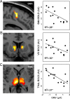
Significant correlation of the CNV amplitude (difference score: monetary reward anticipation > control) and BOLD fMRI signal (monetary reward anticipation > control) within the predefined ROIs (A, SMA; B, VS; C, THAL). The scatterplots show ROI average BOLD signal of all voxels within VS, SMA, and THA ROI. Note that results for CNV1 are shown and that the effects for CNV2 are consistently weaker (data not shown).
Figure 3.
Figure 3.
Mediation analyses. Relation between the EEG CNV and the fMRI response (A) in the VS and (B) in the SMA is fully mediated by the THAL, i.e., the significance of bivariate relations (gray numbers) fully vanishes (red numbers) when THAL thalamus is considered as a mediator. Note that the converse is not true, i.e., the relation between the thalamic fMRI response and the EEG CNV is not significantly reduced when either the VS (C) or the SMA (D) is included as a mediator. Numbers are correlation coefficients; *p < 0.05; **p < 0.01.
Figure 4.
Figure 4.
Results of the DCM analyses. The exceedance probabilities resulting from the BMS for the 32 models are shown in A. The fully connected model is the clear winner (p = 0.71). B, The endogenous connectivity, i.e., connectivity regardless of task demands. C, Modulation of connectivity due to monetary reward anticipation.
Figure 5.
Figure 5.
Partial regression plots showing the relationship of the modulated DCM parameters from SMA→VS (left) and SMA→THAL (right) with the CNV amplitude (difference score: monetary reward anticipation > control) beyond pure THAL BOLD activation effects.

References

    1. Abler B, Erk S, Walter H. Human reward system activation is modulated by a single dose of olanzapine in healthy subjects in an event-related, double-blind, placebo-controlled fMRI study. Psychopharmacology. 2007;191:823–833. doi: 10.1007/s00213-006-0690-y. - DOI - PubMed
    1. Allen PJ, Polizzi G, Krakow K, Fish DR, Lemieux L. Identification of EEG events in the MR scanner: the problem of pulse artifact and a method for its subtraction. Neuroimage. 1998;8:229–239. doi: 10.1006/nimg.1998.0361. - DOI - PubMed
    1. Allen PJ, Josephs O, Turner R. A method for removing imaging artifact from continuous EEG recorded during functional MRI. Neuroimage. 2000;12:230–239. doi: 10.1006/nimg.2000.0599. - DOI - PubMed
    1. Baron RM, Kenny DA. The moderator-mediator variable distinction in social psychological research: conceptual, strategic, and statistical considerations. J Pers Soc Psychol. 1986;51:1173–1182. doi: 10.1037/0022-3514.51.6.1173. - DOI - PubMed
    1. Birbaumer N, Elbert T, Canavan AG, Rockstroh B. Slow potentials of the cerebral cortex and behavior. Physiol Rev. 1990;70:1–41. - PubMed

Publication types

LinkOut - more resources