. 2013 Sep 4;33(36):14558-66.
doi: 10.1523/JNEUROSCI.2001-12.2013.

NG2 cells in white matter but not gray matter proliferate in response to PDGF

Affiliations

NG2 cells in white matter but not gray matter proliferate in response to PDGF

Robert A Hill et al. J Neurosci. .

Abstract

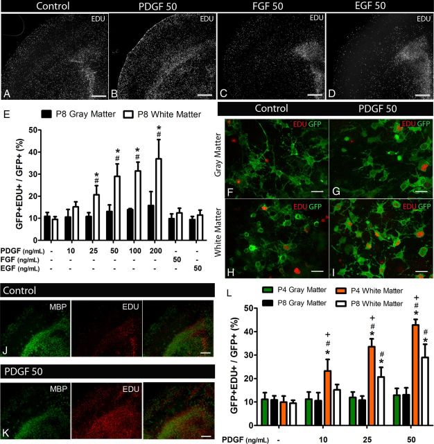
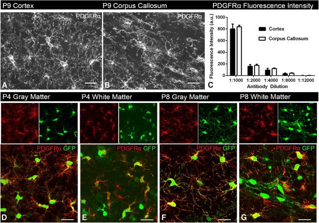
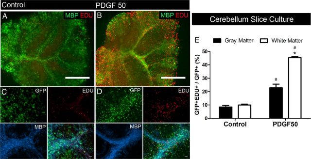
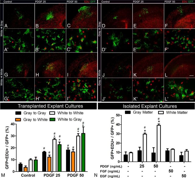
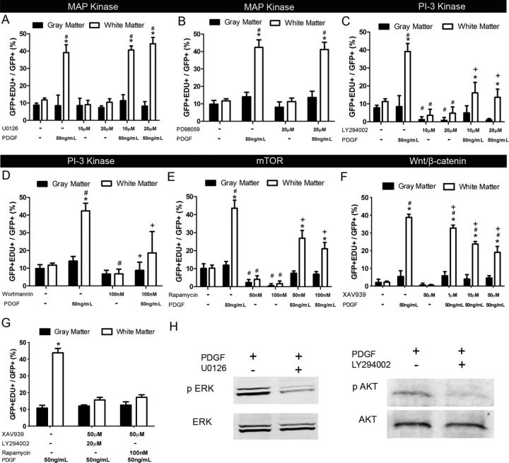
Glial cells that express the NG2 proteoglycan and the α receptor for PDGF (NG2 cells, polydendrocytes) make up the fifth major cell population that serves as oligodendrocyte progenitor cells in the postnatal CNS. Although recent studies have suggested differences in their proliferation and oligodendrocyte differentiation in gray and white matter, the mechanism underlying the observed differences has been unclear. Using organotypic slice cultures from the forebrain and cerebellum of early postnatal NG2creBAC:ZEG mice, we have compared basal and growth factor-induced proliferation of NG2 cells in gray and white matter. NG2 cells in white matter exhibited greater proliferative response to PDGF AA than those in gray matter. Heterotopic slice transplant and explant cultures suggested intrinsic mechanisms for the differential proliferative response of gray and white matter cells. Additionally, younger white matter NG2 cells showed a more robust proliferative response to PDGF. Basal and PDGF-induced proliferation of gray and white matter NG2 cells was largely dependent on Wnt/β-catenin and phosphatidylinositol 3-kinase acting through the mammalian target of rapamycin pathway and not through ERK. These data uncover a previously unrecognized divergence between gray and white matter NG2 cells in the developing brain in their proliferative response to PDGF.


Figures

Figure 1.
Figure 1.
PDGF-induced proliferation of white matter NG2 cells in forebrain slice cultures of NG2creBAC:ZEG mice. Low-magnification images showing EDU+ cells in forebrain slice cultures exposed to no growth factor (A), 50 ng/ml PDGF (B), FGF2 (C), and EGF (D) for 48 h. Quantification of GFP+EDU+ cells in P8 neocortex (gray matter) and corpus callosum (white matter) after exposure to the different growth factors (E) showing increased proportion of proliferating white matter GFP+ cells after exposure to PDGF but not FGF2 or EGF. Dose–response to PDGF demonstrates that gray matter cells do not increase their proliferation even at and above saturating concentrations of PDGF for white matter cells. High-magnification images taken from gray (F, G) and white (H, I) matter regions showing GFP fluorescence and EDU labeling in control (F, H) and PDGF-treated (G, I) slices. MBP and EDU double labeling of cortical slice cultures in control (J) and after exposure to 50 ng/ml PDGF (K) identifies the gray and white matter regions and demonstrates the localization of proliferating EDU+ cells to white matter after PDGF exposure. Quantification of GFP+EDU+ cells in gray and white matter of forebrain slices isolated from P4 and P8 animals (L) demonstrating an age-dependent differential response to PDGF by white matter NG2 cells. Scale bars: A–D, J, K, 100 μm; F–I, 25 μm. *p < 0.001, compared with gray matter (two-way ANOVA, Bonferroni post test). #p < 0.05, compared with same region control (two-way ANOVA, Bonferroni post test). +p < 0.001, compared with P8 (two-way ANOVA, Bonferroni post test). Error bars indicate SD.
Figure 2.
Figure 2.
PDGFRα expression is similar between gray and white matter regions. Images taken from P9 cortex (A) and corpus callosum (B) immunolabeled for PDGFRα. Fluorescence intensity measurements taken from both the cortex and corpus callosum at different antibody concentrations (C) show similar PDGFRα immunofluorescence intensity in both areas, suggesting that PDGFRα expression is similar between gray and white matter (two-way ANOVA, Bonferroni post test). A.U., arbitrary units. Images taken from P4 (D, E) and P8 (F, G) slice cultures from gray and white matter regions of NG2creBAC:ZEG mice immunolabeled for PDGFRα (red), demonstrating similar expression levels of PDGFRα between the two regions and two ages. Green represents GFP fluorescence. Scale bars, 25 μm. Error bars indicate SD.
Figure 3.
Figure 3.
PDGF-induced proliferation of white matter NG2 cells in cerebellar slice cultures. Low-magnification images showing EDU+ cells in the control condition (A) and after 48 h of exposure to 50 ng/ml PDGF (B). MBP immunolabeling (green) shows white matter. Simultaneous detection of GFP, EDU, and MBP (C, D) showing an increase in the proportion of EDU+GFP+ cells in white matter regions (MBP+) in slices exposed to PDGF (D) compared with control (C). E, Quantification showed a significant increase in the proportion of proliferating GFP+ cells in both gray and white matter regions, with a greater magnitude of increase in white matter regions. Scale bars: A, B, 100 μm; C, D, 25 μm. *p < 0.01, compared with gray matter (two-way ANOVA, Bonferroni post test). #p < 0.01, compared with same region control (two-way ANOVA, Bonferroni post test). Error bars indicate SD.
Figure 4.
Figure 4.
NG2 cell proliferation in transplanted and isolated explants. Representative low- (A–L) and high-magnification (A′–L′) images of gray or white matter explants taken from NG2creBAC:ZEG mice and transplanted to gray or white matter regions of wild-type littermate slice cultures. Quantification of transplanted cells (M) shows a greater increase in the proportion of GFP+ cells that were EDU+ originating from the white matter explants when exposed to 25 ng/ml and 50 ng/ml PDGF. Quantification of isolated explants from gray and white matter (N) showed significant increases in the proportion of proliferating GFP+ cells only in white matter explants exposed to PDGF but not FGF2 or EGF. Scale bars: A–L, 100 μm; A′–L′, 25 μm. *p < 0.01, compared with gray matter (two-way ANOVA, Bonferroni post test). #p < 0.01, compared with same region control (two-way ANOVA, Bonferroni post test). Error bars indicate SD.
Figure 5.
Figure 5.
Basal and PDGF-induced proliferation of gray and white matter NG2 cells is dependent on the PI3K–mTOR and Wnt/β-catenin pathways. Quantification of slices exposed to the ERK1/2 inhibitors U0126 (A) and PD98059 (B) showed no change in the proportion of GFP+ cells that were EDU+ with or without PDGF. Quantification of slices exposed to PI3K inhibitors LY294002 (C) and wortmannin (D), the mTOR inhibitor rapamycin (E), and the tankyrase inhibitor XAV939 (F) showed a significant decrease in the proportion of GFP+ cells that were EDU+ in both gray and white matter, even when exposed to PDGF. G, Quantification of slices exposed to XAV939 and either LY294002 or rapamycin further reduced PDGF-induced proliferation in white matter close to the basal level. H, Representative Western blots showing effective inhibition of p44/42 MAPK ERK1/2 phosphorylation by U0126 and Akt S473 phosphorylation by LY294002 in slice cultures in the presence of PDGF. *p < 0.001, compared with gray matter (two-way ANOVA, Bonferroni post test). #p < 0.05, compared with same region control without PDGF (two-way ANOVA, Bonferroni post test). +p < 0.05, compared with 50 ng/ml PDGF only (two-way ANOVA, Bonferroni post test). Error bars indicate SD.

References

    1. Albert M, Antel J, Brück W, Stadelmann C. Extensive cortical remyelination in patients with chronic multiple sclerosis. Brain Pathol. 2007;17:129–138. doi: 10.1111/j.1750-3639.2006.00043.x. - DOI - PMC - PubMed
    1. Azim K, Butt AM. GSK3beta negatively regulates oligodendrocyte differentiation and myelination in vivo. Glia. 2011;59:540–553. doi: 10.1002/glia.21122. - DOI - PubMed
    1. Bahr BA, Kessler M, Rivera S, Vanderklish PW, Hall RA, Mutneja MS, Gall C, Hoffman KB. Stable maintenance of glutamate receptors and other synaptic components in long-term hippocampal slices. Hippocampus. 1995;5:425–439. doi: 10.1002/hipo.450050505. - DOI - PubMed
    1. Baron W, Metz B, Bansal R, Hoekstra D, de Vries H. PDGF and FGF-2 signaling in oligodendrocyte progenitor cells: regulation of proliferation and differentiation by multiple intracellular signaling pathways. Mol Cell Neurosci. 2000;15:314–329. doi: 10.1006/mcne.1999.0827. - DOI - PubMed
    1. Baron W, Shattil SJ, ffrench-Constant C. The oligodendrocyte precursor mitogen PDGF stimulates proliferation by activation of alpha(v)beta3 integrins. EMBO J. 2002;21:1957–1966. doi: 10.1093/emboj/21.8.1957. - DOI - PMC - PubMed

Publication types

MeSH terms

Substances

LinkOut - more resources