. 2013 Sep 4;33(36):14579-90.
doi: 10.1523/JNEUROSCI.2108-13.2013.

Recombinant probes reveal dynamic localization of CaMKIIα within somata of cortical neurons

Affiliations

Recombinant probes reveal dynamic localization of CaMKIIα within somata of cortical neurons

Rudy J Mora et al. J Neurosci. .

Abstract

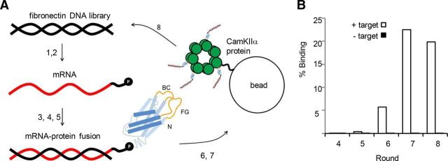
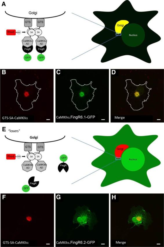
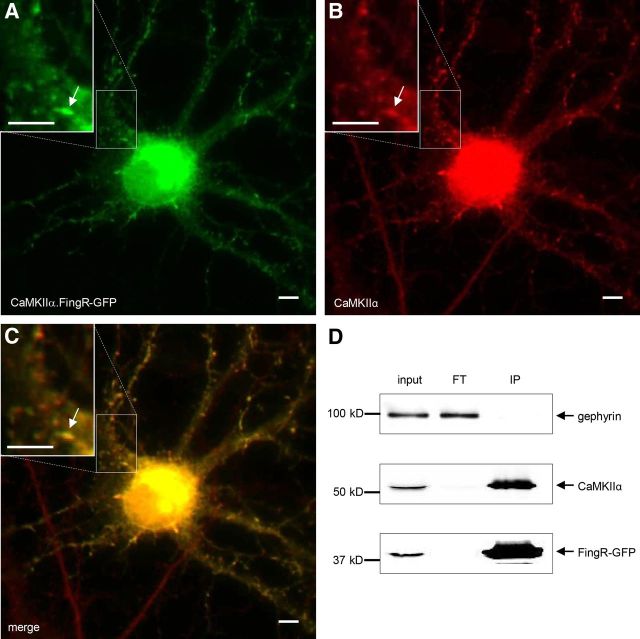
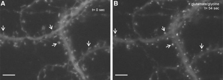
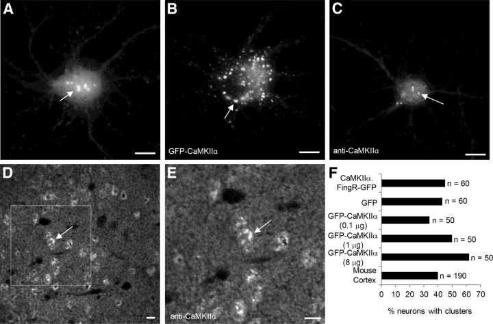
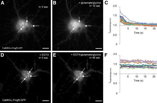
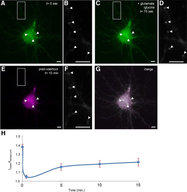
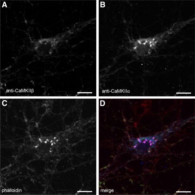
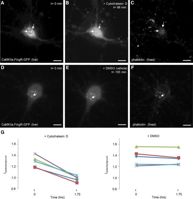
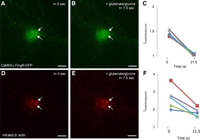
In response to NMDA receptor stimulation, CaMKIIα moves rapidly from a diffuse distribution within the shafts of neuronal dendrites to a clustered postsynaptic distribution. However, less is known about CaMKIIα localization and trafficking within neuronal somata. Here we use a novel recombinant probe capable of labeling endogenous CaMKIIα in living rat neurons to examine its localization and trafficking within the somata of cortical neurons. This probe, which was generated using an mRNA display selection, binds to endogenous CaMKIIα at high affinity and specificity following expression in rat cortical neurons in culture. In ∼45% of quiescent cortical neurons, labeled clusters of CaMKIIα 1-4 μm in diameter were present. Upon exposure to glutamate and glycine, CaMKIIα clusters disappeared in a Ca(2+)-dependent manner within seconds. Moreover, minutes after the removal of glutamate and glycine, the clusters returned to their original configuration. The clusters, which also appear in cortical neurons in sections taken from mouse brains, contain actin and disperse upon exposure to cytochalasin D, an actin depolymerizer. In conclusion, within the soma, CaMKII localizes and traffics in a manner that is distinct from its localization and trafficking within the dendrites.


Figures

Figure 1.
Figure 1.
mRNA display selection on CaMKIIα association domain. A, A round of selection consisting of (1) in vitro transcription of the fibronectin DNA library, (2) ligation of the resulting mRNA (red) to a DNA linker (black) fused to puromycin (P), (3) in vitro translation of mRNA and covalent linkage of fibronectin polypeptide (blue) to puromycin (randomized BC and FG loops in yellow), (4) oligo(dT) purification of mRNA-polypeptide fusions, (5) reverse transcription of mRNA to produce cDNA (black), (6) exposure of the mRNA/DNA-puromycin-polypeptide library to CaMKIIα association domain oligomers immobilized on agarose beads, (7) purification of mRNA/DNA-puromycin-polypeptides that bind at high affinity to CaMKIIα through affinity chromatography, and (8) PCR amplification of purified mRNA/DNA-puromycin-polypeptides to reconstitute a new DNA library of CaMKIIα-specific fibronectins. B, Percentage of mRNA/DNA-puromycin-polypeptides in Rounds 4–7 that bind to either CaMKIIα immobilized on agarose beads (+ target) or to beads alone (− target) in a hot binding assay. The percentage binding to target increases with later rounds, with negligible nonspecific binding.
Figure 2.
Figure 2.
Intracellular Golgi localization assay for FingR/target binding. A, Schematic shows CaMKIIα target targeted to the surface of the Golgi through a Golgi targeting signal (GTS) following expression in COS-7 cells. Coexpressed FingRs that bind to the CaMKIIα target are colocalized with the target at the Golgi. B, C, COS-7 cell coexpressing a Golgi-targeted CaMKIIα association domain (B, GTS-SA-CaMKIIα, red) and a FingR that binds to CaMKIIα (C, CaMKIIα.FingR6.1-GFP, green). D, Both GTS-SA-CaMKIIα and CaMKIIα.FingR6.1-GFP colocalize at the Golgi. E, Schematic of a FingR that does not bind to target following expression in COS-7 cells. F, GTS-SA-CaMKIIα (red) targeted to the Golgi. G, CaMKIIα.FingR6.2-GFP (green) is localized nonspecifically and does not colocalize with GTS-SA-CaMKIIα. H, Merged image shows that CaMKIIα.FingR6.2-GFP and GTS-SA-CaMKIIα do not overlap, indicating that the FingR does not bind at high affinity to the target. Scale bars, 5 μm.
Figure 3.
Figure 3.
CaMKIIα.FingR-GFP colocalizes with endogenous CaMKIIα in neurons. A, B, CaMKIIα.FingR-GFP (A, green) expressed in a dissociated cortical neuron colocalizes similarly to CaMKIIα (B, red). C, Yellow color of merged image of CaMKIIα and CaMKIIα.FingR-GFP indicates colocalization of the two proteins. D, Immunoprecipitation of CaMKIIα.FingR-GFP expressed in cortical neurons in dissociated culture results in coprecipitation of virtually 100% of endogenous CaMKIIα, indicated by its absence in flow through (FT). In contrast, endogenous gephyrin is not coprecipitated. Scale bars, 5 μm.
Figure 4.
Figure 4.
CaMKIIα.FingR-GFP translocates to dendritic spines upon excitatory stimulation. A, CaMKIIα.FingR-GFP expressed in cortical neurons in dissociated culture localizes in a diffuse fashion. B, 54 s after addition of 50 μm glutamate and 5 μm glycine to the bath CaMKIIα.FingR-GFP localizes specifically to puncta corresponding to the tips of dendritic spines. Scale bars, 5 μm.
Figure 5.
Figure 5.
CaMKIIα clusters in the cell bodies of cortical neurons in vitro and in vivo. A, CaMKIIα.FingR-GFP localizes in clusters ∼1–4 μm in diameter in the cell bodies of live cortical neurons. B, GFP-CaMKIIα also clusters in the cell body, but the number of clusters was larger and the intensity more variable than clusters of CaMKIIα.FingR-GFP in live cortical neurons. C, Endogenous CaMKIIα, stained with an antibody is present in clusters in the cell bodies of fixed cortical neurons in culture. D, E, Sections of postnatal day (P) 90 mouse cortex stained with anti-CaMKIIα antibody show clusters within neuronal somata. F, Approximately 45% of cells in dissociated culture showed clusters of CaMKIIα when marked with CaMKIIα.FingR-GFP or with anti-CaMKIIα antibody staining. A similar percentage was found in sections cut from mouse cortex and stained with a CaMKIIα antibody. Although clusters were found in ∼35% of cells transfected with 0.1 μg of CaMKIIα-GFP, increasing the amount of CaMKIIα-GFP caused an increase in the percentage of cells exhibiting clusters, suggesting that expression of the exogenous protein facilitated cluster formation. Approximately 40% of cells in cortices cut from P90 mouse cortex are positive for CaMKIIα clusters. Scale bars, 10 μm.
Figure 6.
Figure 6.
CaMKIIα clusters rapidly disperse upon exposure to high concentrations of Ca2+. A, CaMKIIα.FingR-GFP labels clusters in the cell bodies of cortical neurons in culture. B, Ten seconds following the addition of 50 μm glutamate and 5 μm glycine, the clusters in A have disappeared. C, Time courses of the ratio of fluorescence of CaMKIIα.FingR-GFP at a point within a cluster versus at a point outside of the clusters (Icluster/background) for 10 different cells show that the clusters disappear at similar rates following exposure of cells to 50 μm glutamate and 5 μm glycine. D, Clusters labeled by CaMKIIα.FingR-GFP in cortical neurons in culture in the presence of 5 mm EGTA. E, Following exposure to 50 μm glutamate and 5 μm glycine for 46 s in the presence of 5 mm EGTA, the clusters in D do not change in shape or intensity. F, Time courses of Icluster/background for CaMKIIα.FingR-GFP in 10 different cells shows that clusters do not disperse when exposed to 50 μm glutamate and 5 μm glycine in the presence of 5 mm EGTA. Scale bars, 10 μm.
Figure 7.
Figure 7.
Following dispersal, clusters labeled with CaMKIIα.FingR-GFP re-form in their original positions and shapes. A, Clusters labeled with CaMKIIα.FingR-GFP in a cortical neuron in culture. B, Dendrite from the same neuron as in A shows faint, diffuse staining by CaMKIIα.FingR-GFP. C, Clusters in A dispersed following addition of 50 μm glutamate and 5 μm glycine. D, CaMKIIα.FingR-GFP labeled tight clusters in same dendrite as in B following addition of 50 μm glutamate and 5 μm glycine. E, Following washout of glutamate and glycine and incubation for 10 min, the clusters re-formed in their original positions and shapes. F, Same dendrite as in B and E showed diffuse labeling by CaMKIIα.FingR-GFP following washout of glutamate and glycine. G, Superimposition of the images of clusters before and after dispersal and re-formation confirms that they re-formed in the same locations and configurations as the original clusters. H, Time course of Icluster/background for CaMKIIα.FingR-GFP in the presence (t = 0 to t = 75 s) and absence (t > 75 s) of glutamate and glycine. Error bars represent SEM. Scale bars, 10 μm.
Figure 8.
Figure 8.
CaMKIIα and CaMKIIβ colocalize with F-actin in clusters within the cell body. A, Cluster in the cell body of a cortical neuron in culture stained with anti-CaMKIIβ antibody. B, C, Same cell as in A colabeled with anti-CaMKIIα (B) or with phalloidin (C). D, Merge confirms that expression patterns of CaMKIIα (blue), CaMKIIβ, (green), and actin (red) overlap. Scale bars, 10 μm.
Figure 9.
Figure 9.
Cytochalasin D disruption of F-actin causes CaMKII clusters to disperse. A, CaMKIIα.FingR-GFP-labeled clusters in cell body of a cortical neuron in culture. B, Addition of cytochalasin D (2 μm) for 98 min results in dispersal of the clusters in A. C, Phalloidin staining of cell in B and C verifies that addition of cytochalasin D mediated dispersal of actin clusters. D–F, Clusters in a cortical pyramidal neuron labeled with CaMKIIα.FingR-GFP (D) are not dispersed by DMSO (E), which also leaves clusters of actin filaments intact (F). G, Icluster/background at t = 0 s and t = 21.5 s for CaMKIIα.FingR-GFP following addition of either cytochalasin D or DMSO. Scale bars, 10 μm.
Figure 10.
Figure 10.
CaMKIIα clusters dissociate from F-actin clusters upon stimulation. A, Clusters labeled with CaMKIIα.FingR-GFP (green) in cortical neurons. B, Addition of 50 μm glutamate and 5 μm glycine for 7.5 s results in dispersal of the clusters. C, Icluster/background for CaMKIIα.FingR-GFP shows that the clusters have completely dispersed within 21.5 s. D, Clusters of β actin fused to mKate2 in the same cell as in A. E, Addition of 50 μm glutamate and 5 μm glycine for 7.5 s does not disperse the clusters. F, Time course of Icluster/background for mKate2-β actin during exposure to 50 μm glutamate and 5 μm glycine. Scale bars, 10 μm.

Similar articles

Cited by

References

    1. Andersson AM, Melin L, Bean A, Pettersson RF. A retention signal necessary and sufficient for Golgi localization maps to the cytoplasmic tail of a Bunyaviridae (Uukuniemi virus) membrane glycoprotein. J Virol. 1997;71:4717–4727. - PMC - PubMed
    1. Aronowski J, Grotta JC, Waxham MN. Ischemia-induced translocation of Ca2+/calmodulin-dependent protein kinase II: potential role in neuronal damage. J Neurochem. 1992;58:1743–1753. doi: 10.1111/j.1471-4159.1992.tb10049.x. - DOI - PubMed
    1. Bayer KU, De Koninck P, Leonard AS, Hell JW, Schulman H. Interaction with the NMDA receptor locks CaMKII in an active conformation. Nature. 2001;411:801–805. doi: 10.1038/35081080. - DOI - PubMed
    1. Bayer KU, LeBel E, McDonald GL, O'Leary H, Schulman H, De Koninck P. Transition from reversible to persistent binding of CaMKII to postsynaptic sites and NR2B. J Neurosci. 2006;26:1164–1174. doi: 10.1523/JNEUROSCI.3116-05.2006. - DOI - PMC - PubMed
    1. Coultrap SJ, Bayer KU. CaMKII regulation in information processing and storage. Trends Neurosci. 2012;35:607–618. doi: 10.1016/j.tins.2012.05.003. - DOI - PMC - PubMed

Publication types

LinkOut - more resources