Clinical Trial
. 2013 Oct 18;288(42):30708-30719.
doi: 10.1074/jbc.M113.478685. Epub 2013 Sep 4.

Transforming growth factor-β (TGF-β)-mediated connective tissue growth factor (CTGF) expression in hepatic stellate cells requires Stat3 signaling activation

Affiliations
Clinical Trial

Transforming growth factor-β (TGF-β)-mediated connective tissue growth factor (CTGF) expression in hepatic stellate cells requires Stat3 signaling activation

Yan Liu et al. J Biol Chem. .

Abstract

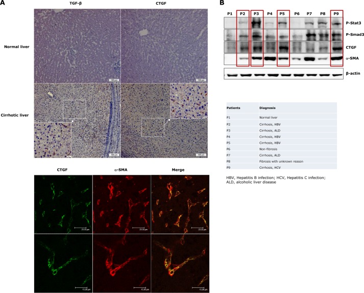
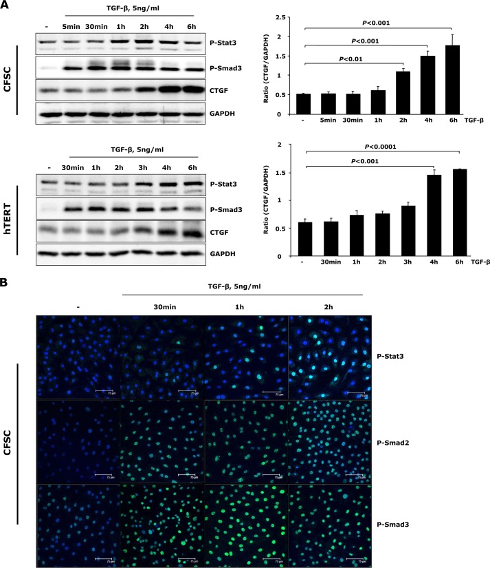
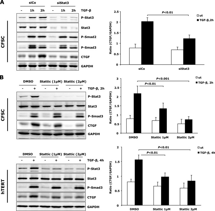
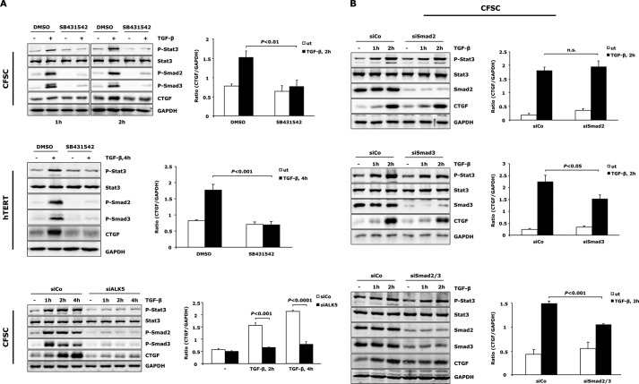
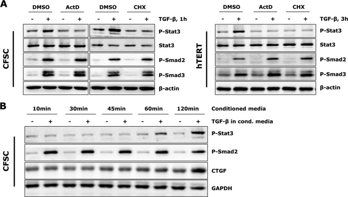
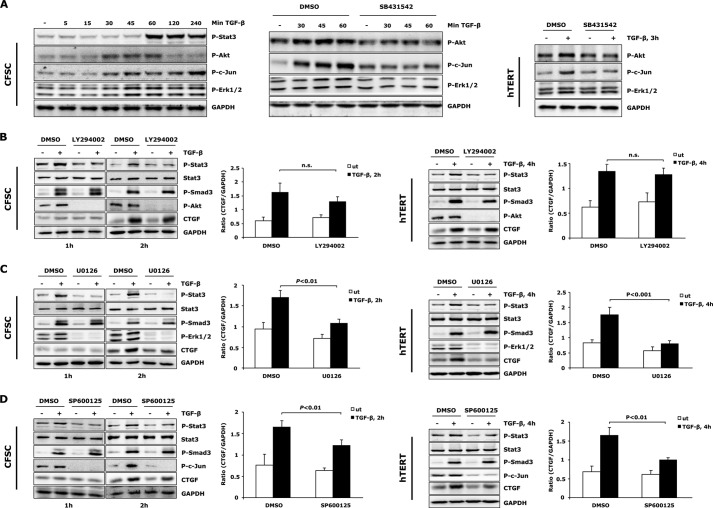
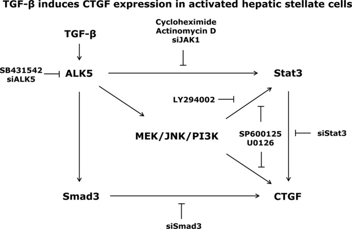
In fibrotic liver, connective tissue growth factor (CTGF) is constantly expressed in activated hepatic stellate cells (HSCs) and acts downstream of TGF-β to modulate extracellular matrix production. Distinct from other cell types in which Smad signaling plays major role in regulating CTGF production, TGF-β stimulated CTGF expression in activated HSCs is only in part dependent on Smad3. Other signaling molecules like MAPKs and PI3Ks may also participate in this process, and the underlying mechanisms have yet to be clarified. In this study, we report involvement of Stat3 activation in modulating CTGF production upon TGF-β challenge in activated HSCs. Stat3 is phosphorylated via JAK1 and acts as a critical ALK5 (activin receptor-like kinase 5) downstream signaling molecule to mediate CTGF expression. This process requires de novo gene transcription and is additionally modulated by MEK1/2, JNK, and PI3K pathways. Cell-specific knockdown of Smad3 partially decreases CTGF production, whereas it has no significant influence on Stat3 activation. The total CTGF production induced by TGF-β in activated HSCs is therefore, to a large extent, dependent on the balance and integration of the canonical Smad3 and Stat3 signaling pathways.

Keywords: ERK; Jak Kinase; Jun N-terminal Kinase (JNK); Liver Fibrosis; MAP Kinases (MAPKs); PI 3-kinase (PI3K); SMAD Transcription Factor.


Figures

FIGURE 1.
FIGURE 1.
Elevated p-Stat3, TGF-β/p-Smad3, and CTGF levels in liver samples of fibrosis/cirrhosis patients. A, immunohistochemical stainings of TGF-β and CTGF expression were performed in healthy liver tissues from a patient with gallstones and in cirrhotic liver tissues from a patient with hepatitis B (HBV) infection (upper panel). Elevated TGF-β and CTGF levels were only detected in cirrhotic liver but not in the liver with normal architecture. Both TGF-β and CTGF displayed positive staining in sinusoidal areas in this patient. Immunofluorescent co-staining demonstrated CTGF overexpression in activated HSCs (α-SMA-positive cells, lower panel). B, liver lysates of fibrosis/cirrhosis patients from different etiologies were analyzed for profibrotic signaling activation and CTGF production. A marked increase of p-Stat3 was detected in association with Smad3 activation and CTGF production in the majority of liver cirrhosis cases (red boxes).
FIGURE 2.
FIGURE 2.
TGF-β induces Stat3 activation and CTGF expression in activated HSCs. A, CFSC-2G (CFSC) cells and hTERT HSCs were treated with 5 ng/ml TGF-β1 for the indicated time periods. Western blot analysis unraveled tyrosine Stat3 phosphorylation prior to significant CTGF expression upon TGF-β challenge. CTGF and GAPDH were quantified by densitometric analysis and expressed as a ratio of CTGF to GAPDH. Data are means ± S.E. p < 0.01, p < 0.001, and p < 0.0001 indicate the statistical significance. B, CFSC-2G cells were stimulated with TGF-β1 for 0.5–2 h. Time-dependent p-Stat3 and p-Smad2/3 nuclear translocation was shown with immunofluorescence.
FIGURE 3.
FIGURE 3.
Stat3 activation is required for TGF-β-mediated CTGF production. A, CFSC-2G (CFSC) cells were transfected with Stat3 or control siRNA for 48 h, followed by exposure to 5 ng/ml TGF-β1 as indicated. Lack of Stat3 resulted in reduced CTGF production. Knockdown was verified at protein level by measurement of total Stat3 and p-Stat3. B, Stattic, a chemical inhibitor, was added to CFSC-2G cells and hTERT HSCs 30 min prior to TGF-β1 treatment to blunt Stat3 phosphorylation. CTGF expression was analyzed by Western blot. Stattic interfered with TGF-β-induced CTGF in a dosage-dependent manner in both cell lines. Data are expressed as a ratio of CTGF to GAPDH and are means ± S.E., p < 0.01 and p < 0.001 indicate the statistical significance. DMSO, dimethyl sulfoxide. ut, untreated; siCo, siControl.
FIGURE 4.
FIGURE 4.
Stat3 activation is downstream of ALK5 and independent of Smad2/3. A, ALK5 is inactivated in CFSC-2G cells and hTERT HSCs by pre-incubation with 5 μm SB431542 for 30 min or by transfection of siRNA for ALK5 in CFSC-2G cells prior to TGF-β1 (5 ng/ml) treatment. Western blot showed repressed Stat3 activation and CTGF expression upon loss of ALK5 activity. Successful inactivation of ALK5 was indicated by reduced p-Smad2/3. B, CFSC-2G cells were transfected with siRNA for Smad2, Smad3, or both, followed by stimulation with 5 ng/ml TGF-β1 as indicated. Knockdown of Smad2 and Smad3 did not interfere with initiation of Stat3 activation. CTGF expression is Smad2-independent but at least in part associated with Smad3 signaling. Results are expressed as a ratio of CTGF to GAPDH and are means ± S.E.; p < 0.05, p < 0.01, and p < 0.001 indicate the statistical significance. DMSO, dimethyl sulfoxide; ut, untreated; siCo, siControl.
FIGURE 5.
FIGURE 5.
TGF-β stimulated Stat3 activation requires de novo protein synthesis. A, CFSC-2G cells and hTERT HSCs were pre-incubated with 1 μg/ml actinomycin D (ActD) or 10 μg/ml cycloheximide (CHX) for 30 min prior to TGF-β1 (5 ng/ml) treatment. TGF-β-induced Stat3 phosphorylation was blocked by both inhibitors. B, CFSC-2G cells were treated with or without TGF-β1 (5 ng/ml) for 1 h, and conditioned media were collected and added into new cell cultures (after overnight starvation). Western blot analysis did not detect earlier Stat3 activation and CTGF expression in conditioned (cond.) media-treated cells. DMSO, dimethyl sulfoxide.
FIGURE 6.
FIGURE 6.
TGF-β-mediated Stat3 phosphorylation requires JAK1. CFSC-2G (CFSC) cells were transfected with JAK1, JAK2, or Tyk2 siRNA and subsequently exposed to 5 ng/ml TGF-β1 for 2 h. Knockdown of JAK1 blunted Stat3 phosphorylation and resulted in reduced CTGF expression. Silencing JAK2 and Tyk2 had no influence on this process. CTGF and GAPDH expression was quantified, and data are means ± S.E.; p < 0.01 indicates the statistical significance. Knockdown of JAKs was verified at the RNA level by semiquantitative PCR. ut, untreated; siCo, siControl; n.s., not significant.
FIGURE 7.
FIGURE 7.
MAPKs and PI3K are required for TGF-β-induced Stat3 activation in activated HSCs. A, Western blot revealed ALK5 (SB431542, 5 μm) mediated activation of Erk1/2, JNK, and PI3K/Akt pathways prior to Stat3 phosphorylation upon TGF-β treatment. B–D, PI3K/Akt (LY294002, 10 μm), Erk1/2 (U0126, 10 μm), and JNK (SP600125, 5 μm) inhibitors were used to block specific signaling pathways. CFSC-2G cells and hTERT HSCs were then treated with TGF-β1 (5 ng/ml) for indicated times to detect Stat3 activation and CTGF expression. All inhibitor treatments attenuated early Stat3 phosphorylation. CTGF expression was reduced by Erk1/2 and JNK inhibition but remained unchanged in the absence of PI3K. In contrast to the enhanced p-Smad3 level in LY294002 treated CFSC-2G cells (TGF-β1, 2 h), a reduced p-Smad3 level was observed in hTERT HSCs after PI3K inhibition (TGF-β1, 4 h), indicating a third signaling pathway participating in modulation of total CTGF production in this setting (see “Discussion”). CTGF and GAPDH expression was densitometrically quantified, and results are means ± S.E., p < 0.01 and p < 0.001 indicate the statistical significance. ut, untreated; DMSO, dimethyl sulfoxide.
FIGURE 8.
FIGURE 8.
Possible mechanism for TGF-β-mediated CTGF expression in activated HSCs. TGF-β binding to ALK5 receptor induces direct Smad phosphorylation and indirect JAK/Stat3 activation, resulting in enhanced CTGF expression in activated HSCs. This process is additionally modulated by ALK5 mediated activation of MEK1/2, JNK, and PI3K pathways. The total amount of CTGF production upon TGF-β stimulation thus results from integration of both Smad3 and Stat3 signaling pathways.

Similar articles

Cited by

References

    1. Leask A., Abraham D. J. (2003) The role of connective tissue growth factor, a multifunctional matricellular protein, in fibroblast biology. Biochem. Cell Biol. 81, 355–363 - PubMed
    1. Lipson K. E., Wong C., Teng Y., Spong S. (2012) CTGF is a central mediator of tissue remodeling and fibrosis and its inhibition can reverse the process of fibrosis. Fibrogenesis Tissue Repair 5, Suppl. 1, S24. - PMC - PubMed
    1. Gressner O. A., Gressner A. M. (2008) Connective tissue growth factor: a fibrogenic master switch in fibrotic liver diseases. Liver Int. 28, 1065–1079 - PubMed
    1. Williams E. J., Gaça M. D., Brigstock D. R., Arthur M. J., Benyon R. C. (2000) Increased expression of connective tissue growth factor in fibrotic human liver and in activated hepatic stellate cells. J. Hepatol. 32, 754–761 - PubMed
    1. Abou-Shady M., Friess H., Zimmermann A., di Mola F. F., Guo X. Z., Baer H. U., Büchler M. W. (2000) Connective tissue growth factor in human liver cirrhosis. Liver 20, 296–304 - PubMed

Publication types

MeSH terms