. 2013 Oct 11;288(41):29654-69.
doi: 10.1074/jbc.M113.504639. Epub 2013 Sep 4.

Inside-out signaling promotes dynamic changes in the carcinoembryonic antigen-related cellular adhesion molecule 1 (CEACAM1) oligomeric state to control its cell adhesion properties

Affiliations

Inside-out signaling promotes dynamic changes in the carcinoembryonic antigen-related cellular adhesion molecule 1 (CEACAM1) oligomeric state to control its cell adhesion properties

Prerna C Patel et al. J Biol Chem. .

Abstract

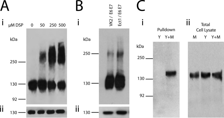
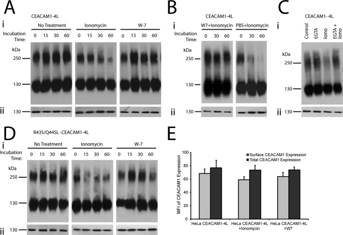
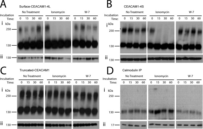
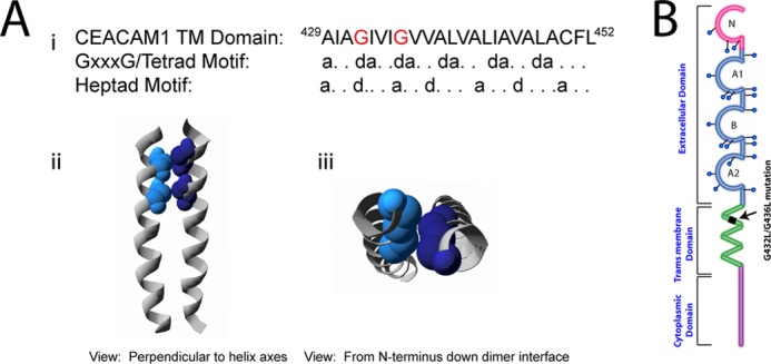
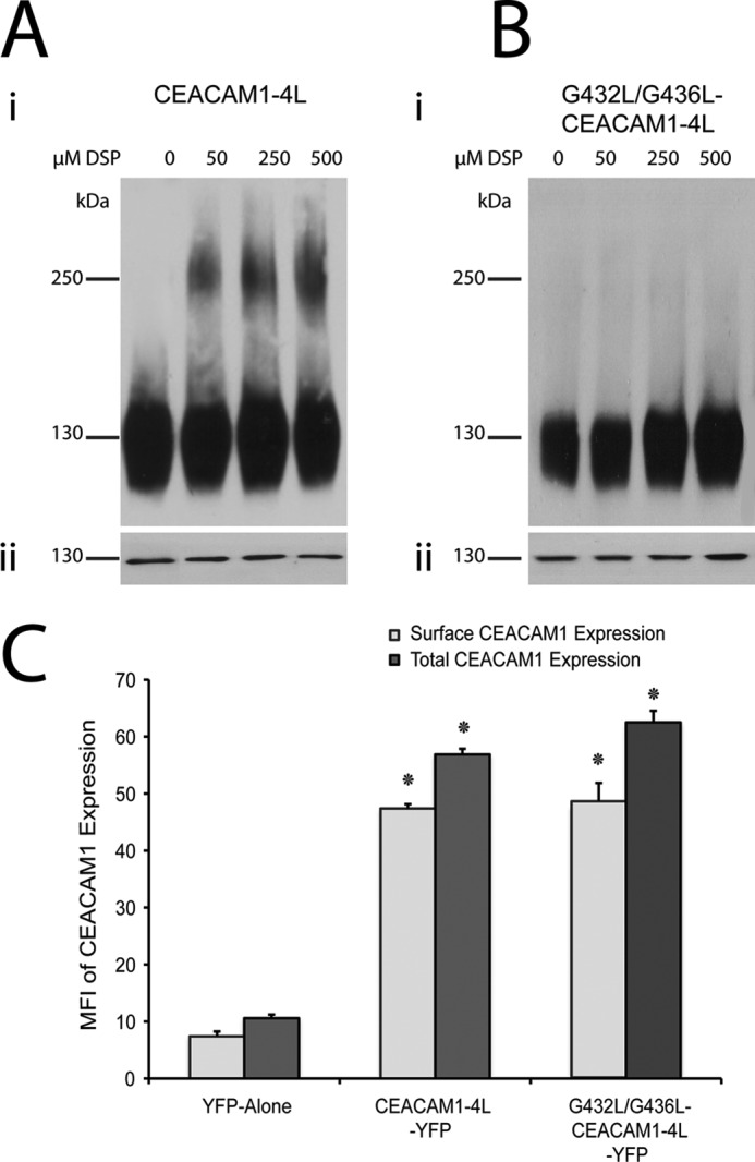
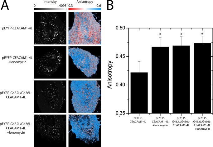
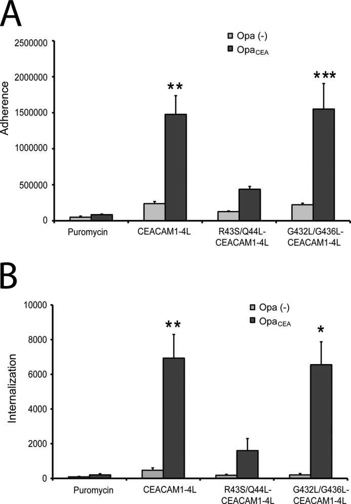
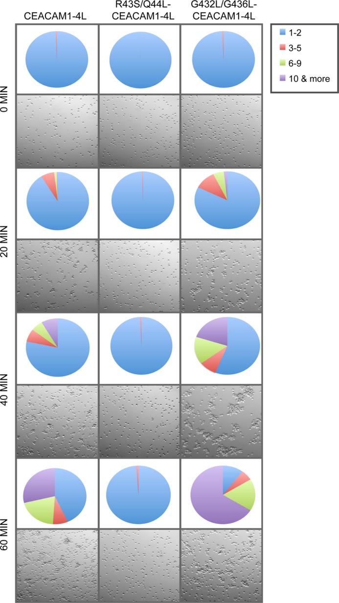
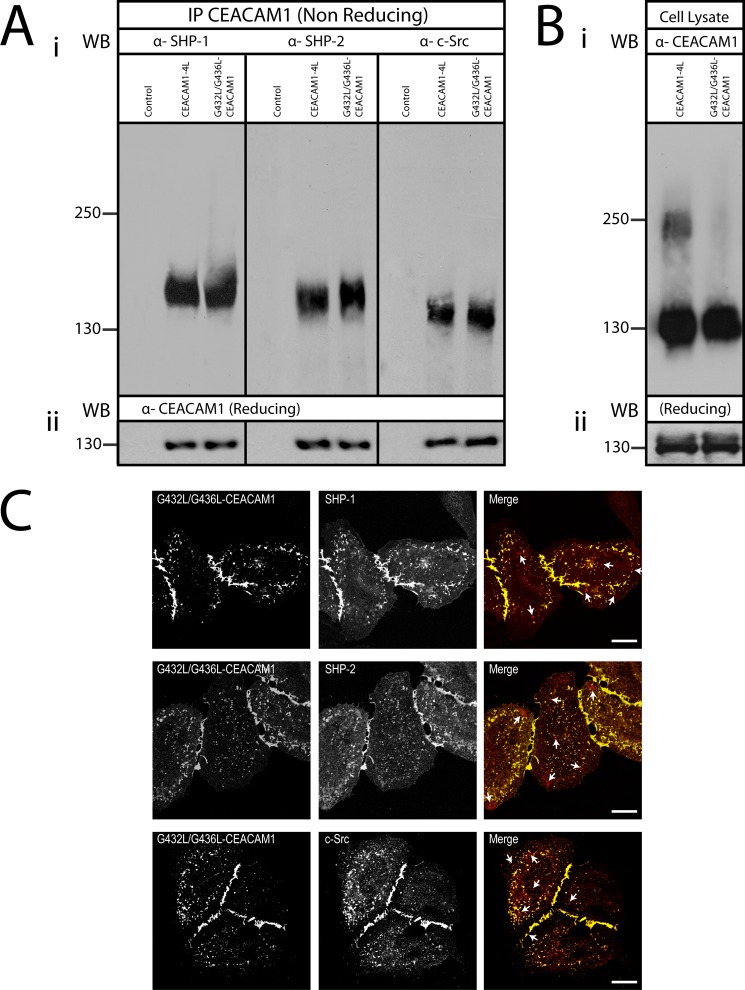
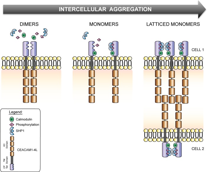
Cell-cell contacts are fundamental to multicellular organisms and are subject to exquisite levels of control. The carcinoembryonic antigen-related cell adhesion molecule 1 (CEACAM1) can engage in both cis-homophilic (parallel) oligomerization and trans-homophilic (anti-parallel) binding. In this study, we establish that the CEACAM1 transmembrane domain has a propensity to form cis-dimers via the transmembrane-embedded (432)GXXXG(436) motif and that this basal state is overcome when activated calmodulin binds to the CEACAM1 cytoplasmic domain. Although mutation of the (432)GXXXG(436) motif reduced CEACAM1 oligomerization, it did not affect surface localization of the receptor or influence CEACAM1-dependent cellular invasion by the pathogenic Neisseria. The mutation did, however, have a striking effect on CEACAM1-dependent cellular aggregation, increasing both the kinetics of cell-cell association and the size of cellular aggregates formed. CEACAM1 association with tyrosine kinase c-Src and tyrosine phosphatases SHP-1 and SHP-2 was not affected by the (432)GXXXG(436) mutation, consistent with their association with the monomeric form of wild type CEACAM1. Collectively, our results establish that a dynamic oligomer-to-monomer shift in surface-expressed CEACAM1 facilitates trans-homophilic binding and downstream effector signaling.

Keywords: Bacterial Adhesion; CEACAM1; Calcium Signaling; Calmodulin; Cancer; Carcinoembryonic Antigen-related Cell Adhesion Molecule; Cell Adhesion; Homodimer; Homophilic Binding; Membrane Proteins.


Figures

FIGURE 1.
FIGURE 1.
CEACAM1 exists as monomers and oligomers in HeLa cells. A, HeLa-CEACAM1 cells were treated with the indicated concentrations of DSP; total cell lysates were resolved under nonreducing (panel i) or reducing (panel ii) conditions by SDS-PAGE and probed with antibodies specific for CEACAM1. B, ectocervical (Ect1/E6E7) and vaginal (Vk2/E6E7) epithelial cells were cross-linked with DSP. Total cell lysates were resolved under nonreducing (panel i) or reducing (panel ii) conditions by SDS-PAGE, and immunoblots were probed with antibodies specific for CEACAM1. C, HeLa cells were transfected with only pEYFP-CEACAM1–4L (Y) or pEYFP-CEACAM1–4L and CEACAM1–4L-c-Myc (Y+M) and treated with DSP. c-Myc-containing proteins were immunoprecipitated and SDS-PAGE immunoblots run in reducing conditions were probed with antibodies specific for YFP (C, panel i). Cell lysates were probed with anti-CEACAM1 antibody to ensure all cells were expressing the transfected proteins (C, panel ii). Blots are representative of three independent experiments.
FIGURE 2.
FIGURE 2.
CEACAM1 dimers are formed by cis-interactions and regulated by intracellular Ca2+. Transfected HeLa cells expressing CEACAM1–4L (A–C) or CEACAM1–4L containing R43S/Q44L mutations (D) were either untreated or treated with ionomycin or W-7 or W-7 and ionomycin or PBS + ionomycin, EGTA, and EGTA + ionomycin for various time points, and then proteins were cross-linked with DSP. Ionomycin was used at 10 μm for B but used at 1 μm for all other experiments. Total cell lysates were resolved under nonreducing (panel i) or reducing (panel ii) conditions by SDS-PAGE, and immunoblots were probed with antibodies specific for CEACAM1. Depicted blots are representative of three independent experiments. E, quantitative evaluation of surface and total CEACAM1 expression using flow cytometry, plotted as mean fluorescence intensity (MFI). Data represent mean ± S.E. from three separate experiments.
FIGURE 3.
FIGURE 3.
Cell surface CEACAM1 dimers are dissociated by Ca2+-calmodulin binding. Transfected HeLa cells expressing full-length CEACAM1 (CEACAM1–4L) were either untreated or treated with ionomycin or W-7, and then proteins were cross-linked with DSP. Cell surface-exposed CEACAM1 was labeled by conjugation to biotin and recovered using streptavidin-agarose beads; biotinylated lysates were resolved under nonreducing (A, panel i) or reducing (A, panel ii) conditions by SDS-PAGE, and immunoblots were probed with antibodies specific for CEACAM1. Transfected HeLa cells expressing a natural splice variant of CEACAM1 containing a short cytoplasmic domain, 10 amino acids (CEACAM1–4S) (B), or a recombinant form completely lacking a cytoplasmic domain (truncated CEACAM1) (C) were either untreated or treated with ionomycin or W-7, and then proteins were cross-linked with DSP. D, co-immunoprecipitation (IP) of HeLa cells expressing full-length CEACAM1–4L using calmodulin-specific antibody after the treatment with or without ionomycin or W-7 and DSP. Calmodulin-associated proteins were resolved under nonreducing conditions, and immunoblots were probed with antibodies specific for CEACAM1 (panel i) or separated under reducing conditions, and immunoblots were probed to detect total levels of calmodulin (panel ii). Blots are representative of three independent experiments.
FIGURE 4.
FIGURE 4.
Computational model of the CEACAM1 transmembrane domain dimers. A, sequence of the human CEACAM1 transmembrane domain is shown. The position of two glycine residues (red text) is consistent with the GXXXG/tetrad motif repeat patterning (at position d) and with heptad repeat motif patterning (at positions d and a) (panel i). A view perpendicular to the helix axes and from the N terminus down the interface of a representative left-handed TMD dimer model is shown. Individual helices are represented as gray ribbons, and the glycine residues at the interface are depicted as space-filling spheres in shades of blue (panels ii and iii). Note the close packing of Gly-432 and Gly-436 in the contact surface between the two helices. B, schematic of CEACAM1 to illustrate location of transmembrane domain mutations of glycine 432 and glycine 436 to Leu residues.
FIGURE 5.
FIGURE 5.
Oligomerization of wild type CEACAM1 and its G432L/G436L transmembrane mutant. Transiently transfected HeLa cells expressing CEACAM1–4L (A) or CEACAM1–4L containing G432L/G436L mutations (B) were treated with various concentrations of the homobifunctional chemical cross-linker DSP; total cell lysates were resolved under nonreducing conditions (panel i) or reducing conditions (panel ii) by SDS-PAGE and probed with antibodies specific for CEACAM1. Blots are representative of three independent experiments. B, quantitative evaluation of CEACAM1 surface as well as total expression by flow cytometry, plotted as mean fluorescence intensity (MFI). The x axis indicates transfected allele. Data represent the mean ± S.E. from three separate experiments. *, p value < 0.05 compared with pEYFP alone control.
FIGURE 6.
FIGURE 6.
FAIM reveals varied oligomerization of CEACAM1–4L and G432L/G436L-CEACAM1–4L. Fluorescence anisotropy measurements were made on HeLa cells expressing enhanced YFP-CEACAM1–4L and enhanced YFP-G432L/G436L-CEACAM1–4L with and without ionomycin treatment. A, intensity images (left column) show similar expression and distribution of wild type and mutant CEACAM1 on the plasma membrane. A comparison of the anisotropy images (right column) show that ionomycin treatment increases the anisotropy of CEACAM1–4L-YFP, indicating that there is an increase in the proportion of CEACAM1 that is in monomeric form. Anisotropy images for the untreated G432L/G436L mutant reflect that seen with ionomycin-treated CEACAM1, and this is not affected by exposure to ionomycin. B, summarized anisotropy values for CEACAM1–4L-YFP and G432L/G436L-CEACAM1–4L-YFP with and without ionomycin. The data represent the mean ± S.E. from measurements on 30 or more cells collected from six independent experiments. Asterisk indicates a p value of <0.05 by a Student's t test compared with the CEACAM1–4L-YFP.
FIGURE 7.
FIGURE 7.
Effects of CEACAM1 dimerization on adhesion and engulfment of N. gonorrhoeae. HeLa cells transfected with vector control, CEACAM1–4L, CEACAM1–4L containing R43S/Q44L mutations, or CEACAM1–4L containing G432L/G436L mutations were infected with N. gonorrhoeae expressing either no Opa proteins (gray bars) or a CEACAM1-specific OpaCEA protein (black bars). Means ± S.E. of triplicate samples were graphed to illustrate bacterial attachment (A) and internalization (B) by these cells. Asterisk denotes p < 0.05; double asterisk, p < 0.01; and triple asterisk, p < 0.005 compared with the untreated samples. The results are representative of three independent experiments.
FIGURE 8.
FIGURE 8.
Effects of CEACAM1 dimerization on trans-homophilic cell-cell adhesion. HeLa cells transfected with CEACAM1–4L, R43S/Q44L-CEACAM1–4L, or G432L/G436L-CEACAM1–4L were prepared as single cell suspensions and then allowed to aggregate. Images of cell cultures taken at indicated time points are shown. Quantitative analysis of number and size of aggregates at each time point are represented as a pie chart. Each value is a mean of triplicate samples and are representative of three independent experiments.
FIGURE 9.
FIGURE 9.
Association of downstream effector proteins with CEACAM1 monomers. A, HeLa cells untransfected (control) or transfected with CEACAM1–4L or G432L/G436L-CEACAM1–4L were pretreated with pervanadate and then cross-linked with DSP. CEACAM1 and associated proteins were co-immunoprecipitated (IP) with CEACAM1-specific antibodies. Proteins were resolved under nonreducing conditions, and immunoblots were probed with antibodies specific for SHP-1, SHP-2, and c-Src (panel i) or separated under reducing conditions and immunoblots probed to detect total levels of CEACAM1 (panel ii). WB, Western blot. B, cell lysates were probed with anti-CEACAM1 antibody to ensure the cells were expressing the transfected proteins under nonreducing (panel i) or reducing conditions (panel ii). Note that CEACAM1-effector complexes migrate at sizes greater than CEACAM1 alone (compare A with B). C, immunostaining of SHP-1, SHP-2 phosphatases, and c-Src kinase in HeLa cells expressing pEYFP-G432L/G436L-CEACAM1–4L. The cellular localization of pEYFP-G432L/G436L-CEACAM1–4L (green false-color YFP) with SHP-1 (red), with SHP-2 (red), or with c-Src (red), as indicated, were analyzed by confocal microscopy. Arrows indicate an area devoid of CEACAM1. Bars, 10 μm. Data are representative of three independent experiments.
FIGURE 10.
FIGURE 10.
Dynamic reassortment of CEACAM1 oligomers to facilitate trans-homophilic binding and downstream effector recruitment. A schematic diagram depicts the progression of CEACAM1 oligomeric states in a basal state (dimers), as monomers bound to Ca2+-calmodulin, and in a lattice-like arrangement at cell-cell contacts.

Similar articles

Cited by

References

    1. Okegawa T., Pong R. C., Li Y., Hsieh J. T. (2004) The role of cell adhesion molecule in cancer progression and its application in cancer therapy. Acta Biochim. Pol. 51, 445–457 - PubMed
    1. Beauchemin N., Draber P., Dveksler G., Gold P., Gray-Owen S., Grunert F., Hammarström S., Holmes K. V., Karlsson A., Kuroki M., Lin S. H., Lucka L., Najjar S. M., Neumaier M., Obrink B., Shively J. E., Skubitz K. M., Stanners C. P., Thomas P., Thompson J. A., Virji M., von Kleist S., Wagener C., Watt S., Zimmermann W. (1999) Redefined nomenclature for members of the carcinoembryonic antigen family. Exp. Cell Res. 252, 243–249 - PubMed
    1. Barnett T. R., Drake L., Pickle W., 2nd (1993) Human biliary glycoprotein gene: characterization of a family of novel alternatively spliced RNAs and their expressed proteins. Mol. Cell Biol. 13, 1273–1282 - PMC - PubMed
    1. Hinoda Y., Neumaier M., Hefta S. A., Drzeniek Z., Wagener C., Shively L., Hefta L. J., Shively J. E., Paxton R. J. (1988) Molecular cloning of a cDNA coding biliary glycoprotein I: primary structure of a glycoprotein immunologically cross-reactive with carcinoembryonic antigen. Proc. Natl. Acad. Sci. U.S.A. 85, 6959–6963 - PMC - PubMed
    1. Barnett T. R., Kretschmer A., Austen D. A., Goebel S. J., Hart J. T., Elting J. J., Kamarck M. E. (1989) Carcinoembryonic antigens: alternative splicing accounts for the multiple mRNAs that code for novel members of the carcinoembryonic antigen family. J. Cell Biol. 108, 267–276 - PMC - PubMed

Publication types

MeSH terms

LinkOut - more resources