. 2013 Oct 18;288(42):30236-30245.
doi: 10.1074/jbc.M113.480079. Epub 2013 Sep 4.

Increased expression of reticulon 3 in neurons leads to reduced axonal transport of β site amyloid precursor protein-cleaving enzyme 1

Affiliations

Increased expression of reticulon 3 in neurons leads to reduced axonal transport of β site amyloid precursor protein-cleaving enzyme 1

Minzi Deng et al. J Biol Chem. .

Abstract

BACE1 is the sole enzyme responsible for cleaving amyloid precursor protein at the β-secretase site, and this cleavage initiates the generation of β-amyloid peptide (Aβ). Because amyloid precursor protein is predominantly expressed by neurons and deposition of Aβ aggregates in the human brain is highly correlated with the Aβ released at axonal terminals, we focused our investigation of BACE1 localization on the neuritic region. We show that BACE1 was not only enriched in the late Golgi, trans-Golgi network, and early endosomes but also in both axons and dendrites. BACE1 was colocalized with the presynaptic vesicle marker synaptophysin, indicating the presence of BACE1 in synapses. Because the excessive release of Aβ from synapses is attributable to an increase in amyloid deposition, we further explored whether the presence of BACE1 in synapses was regulated by reticulon 3 (RTN3), a protein identified previously as a negative regulator of BACE1. We found that RTN3 is not only localized in the endoplasmic reticulum but also in neuritic regions where no endoplasmic reticulum-shaping proteins are detected, implicating additional functions of RTN3 in neurons. Coexpression of RTN3 with BACE1 in cultured neurons was sufficient to reduce colocalization of BACE1 with synaptophysin. This reduction correlated with decreased anterograde transport of BACE1 in axons in response to overexpressed RTN3. Our results in this study suggest that altered RTN3 levels can impact the axonal transport of BACE1 and demonstrate that reducing axonal transport of BACE1 in axons is a viable strategy for decreasing BACE1 in axonal terminals and, perhaps, reducing amyloid deposition.

Keywords: Alzheimer Disease; Amyloid Precursor Protein; Axonal Transport; BACE1; Confocal Microscopy; Kymograph; RTN3; Secretases; Synapses; Synaptic Localization.


Figures
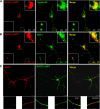
FIGURE 1.
FIGURE 1.
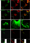
Neuronal localization of BACE1. Cultured mouse hippocampal neurons at DIV7 were transfected with either BACE1-mRFP (A and B) or BACE1-eGFP (C) for 48 h. The fixed cultured neurons were immunostained with an antibody specific to syntaxin 6 to label late Golgi and TGN compartments (A), EEA1 for early endosomal (B), or MAP2 for dendrites (C). The boxed area is enlarged and shown as an inset.
FIGURE 2.
FIGURE 2.
Neuronal localization of RTN3. The RTN3-eGFP expression construct was used to infect cultured mouse hippocampal neurons for 48 h, and the fixed neurons were reacted with anti-calnexin to mark the ER (A) or MAP2 to mark dendrites (F). The Sec61-myc expression construct (B) was coexpressed with RTN3 to determine their colocalization. C and D, the RTN3-eGFP expression construct was used to transfect endothelial cells, and fixed cells were visualized and imaged using a confocal microscope. The images showed clear localization of RTN3 in the tubular ER. The boxed area is enlarged and shown as an inset. E, higher magnification of the ER structure of cultured neurons transfected with the RTN3-eGFP expression construct was captured to observe tubular ER in neurons. Scale bars = 2.5 μm in E and 10 μm in the other panels.
FIGURE 3.
FIGURE 3.
RTN3 colocalizes with BACE1 in the soma. Cultured mouse neurons were coinfected with BACE1-mRFP or RTN3-eGFP for 48 h, and fixed neurons were imaged by a confocal microscope. Colocalization of BACE1 with RTN3 was mainly in the soma (A). BACE1 in axons were significantly weaker than RTN3 (A), and partial colocalization of these two proteins is evident in B. The boxed area is enlarged and shown as an inset. Scale bar = 10 μm.
FIGURE 4.
FIGURE 4.
Colocalization of RTN3 with synaptophysin. A and B, cultured mouse hippocampal neurons were infected with a lentiviral vector expressing either eGFP or RTN3-eGFP for 48 h, and fixed neurons were then reacted with an antibody specific to synaptophysin, a protein abundantly enriched in presynaptic vesicles. C, RTN3-myc and eGFP were cotransfected in cultured neurons, and staining of dendritic spines was labeled by eGFP. The boxed area is enlarged and shown as an inset. Scale bars = 10 μm in A and B.
FIGURE 5.
FIGURE 5.
RTN3 reduces colocalization of BACE1 with synaptophysin. Cultured neurons were infected with the BACE1-mRFP lentivirus-expressing construct together with either lentiviral vector expressing eGFP (A) or RTN3-eGFP (B) for 48 h. After transfection, neurons were fixed and examined for BACE1 colocalization with synaptophysin. C, an enlarged view was captured to determine BACE1-mRFP in spines. The boxed area is enlarged and shown as an inset. Scale bars = 10 μm and 7.5 μm in C.
FIGURE 6.
FIGURE 6.
Quantitative analysis of the colocalization of BACE1 with synaptophysin. Neurons were infected with BACE1-mRFP together with a vector expressing either control eGFP (A) or RTN3-eGFP (B). BACE1-mRFP fluorescent particles were selected randomly. Line profiles were plotted on merged images for the same BACE1 particles (lower panels). The x axis represents the distance along the line, and the y axis represents the pixel intensity. Colocalization of BACE1 and synaptophysin at each point was defined as overlapping red and blue peaks. C, a total of 118 BACE1-mRFP fluorescent particles from 20 axons were analyzed for colocalization with synaptophysin. Results are summarized from three independent experiments. **, p = 0.0014; Student's t test.
FIGURE 7.
FIGURE 7.
Overexpression of RTN3 reduces BACE1 particles scattered along the neurites. Neurons were infected with the indicated lentiviral expression constructs, and neurons with coexpression of BACE1-mRFP and eGFP (A) or BACE1-mRFP and RTN3-eGFP (B) were analyzed. The images of neurites from the corresponding neurons were enlarged and are presented in the lower panels. Scale bar = 25 μm. C, a statistical analysis of a total of 20 neurites from three independent experiments was conducted. The number of BACE1 particles in BACE1-mRFP/eGFP (N1) coinfected neurons was decreased from 1.28 particles/unit length in the neurites to 0.62 particles in the RTN3 overexpression condition. ***, p = 0.0005; Student's t test with Welch's correction. D, analysis of BACE1-eGFP puncta size in the neuronal somata was performed by measuring the average size of the five biggest aggregates in one neuron, and a total of 25 neurons from three independent experiments were chosen for quantification. **, p = 0.0015; Student's t test with Welch's correction.
FIGURE 8.
FIGURE 8.
Reducing anterograde transport of BACE1 in axons by overexpressed RTN3. A, representative kymographs of BACE1-eGFP-containing vesicles in the axon. Cultured neurons at DIV7 were transfected with BACE1-eGFP together with either pcDNA3.1 empty vector (3.1) or RTN3-myc for 48 h. Live cells were imaged at 1 s/interval for 1 min using confocal microscopy with ×630 magnification. Kymographs of BACE1-positive vesicles trafficking along axons were analyzed by ImageJ. Right or left descending particles represent anterograde or retrograde transport in axons. B–D, axonal transport of BACE1-containing vesicles was quantitatively compared with or without RTN3 overexpression. Flux rates of BACE1-eGFP-containing vesicles were measured. Of three independent experiments, a total of 457 BACE1-eGFP vesicles from 22 RTN3 cotransfected neurons and 583 vesicles from 22 neurons transfected with BACE1-eGFP and empty vector were quantified. **, p = 0.0014; Student's t test. E and F, the trafficking rate of BACE1-eGFP vesicles in either the anterograde or retrograde direction was calculated. Anterograde trafficking of BACE 1 was decreased from 15.85 μm/min to 6.68 μm/min in the presence of overexpressed RTN3. ***, p < 0.0001; n = 20 cells; Student's t test with Welch's correction (E). On the other hand, trafficking of BACE1-eGFP vesicles was not significantly affected in the anterograde direction (F).
FIGURE 9.
FIGURE 9.
RTN3 has no effects on axonal transport of APP. A, representative kymographs of APP-eGFP-containing vesicles in the axon. The Swedish mutant version of APP was fused to eGFP to generate APP-eGFP, which was transfected together with either RTN3-myc or its empty vector into neurons at DIV7 for about 48 h. Cells were then imaged at 1 s/interval for 1 min. B–D, RTN overexpression does not significantly alter the percentage of stationary or mobile APP-eGFP vesicles. A total of 515 APP-eGFP vesicles from 20 neurons cotransfected with APP and RTN3 and 516 vesicles from 20 neurons transfected with APP-eGFP and empty vector were quantified to determine the percentage of vesicles either being transported or stationary (17.13 ± 2.67% versus 18.93 ± 1.77% in anterograde transport; 15.13 ± 2.02% versus 15.61 ± 2.31% in retrograde transport; 67.74 ± 3.98% versus 65.47 ± 3.28% stationary; **, p > 0.05, Student's t test.

Similar articles

Cited by

References

    1. Selkoe D. J., Yamazaki T., Citron M., Podlisny M. B., Koo E. H., Teplow D. B., Haass C. (1996) The role of APP processing and trafficking pathways in the formation of amyloid β-protein. Ann. N.Y. Acad. Sci. 777, 57–64 - PubMed
    1. Sisodia S. S., St George-Hyslop P. H. (2002) γ-Secretase, Notch, Aβ and Alzheimer's disease. Where do the presenilins fit in? Nat. Rev. Neurosci. 3, 281–290 - PubMed
    1. Haass C. (2004) Take five. BACE and the γ-secretase quartet conduct Alzheimer's amyloid β-peptide generation. EMBO J. 23, 483–488 - PMC - PubMed
    1. Hussain I., Powell D., Howlett D. R., Tew D. G., Meek T. D., Chapman C., Gloger I. S., Murphy K. E., Southan C. D., Ryan D. M., Smith T. S., Simmons D. L., Walsh F. S., Dingwall C., Christie G. (1999) Identification of a novel aspartic protease (Asp 2) as β-secretase. Mol. Cell Neurosci. 14, 419–427 - PubMed
    1. Vassar R., Bennett B. D., Babu-Khan S., Kahn S., Mendiaz E. A., Denis P., Teplow D. B., Ross S., Amarante P., Loeloff R., Luo Y., Fisher S., Fuller J., Edenson S., Lile J., Jarosinski M. A., Biere A. L., Curran E., Burgess T., Louis J. C., Collins F., Treanor J., Rogers G., Citron M. (1999) β-Secretase cleavage of Alzheimer's amyloid precursor protein by the transmembrane aspartic protease BACE. Science 286, 735–741 - PubMed

Publication types

MeSH terms