. 2013 Sep 3;14(9):18009-23.
doi: 10.3390/ijms140918009.

Celf1 is required for formation of endoderm-derived organs in zebrafish

Affiliations

Celf1 is required for formation of endoderm-derived organs in zebrafish

Naoyuki Tahara et al. Int J Mol Sci. .

Abstract

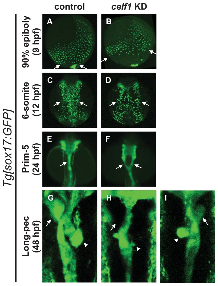
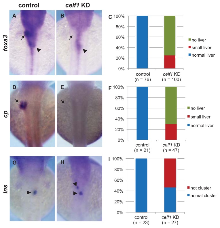
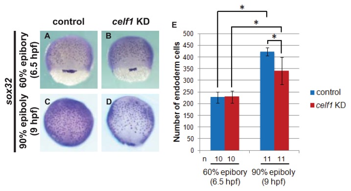
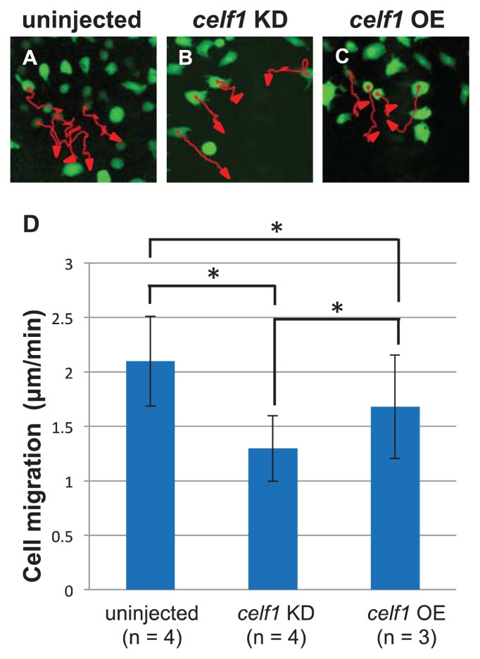
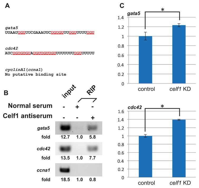
We recently reported that an RNA binding protein called Cugbp Elav-like family member 1 (Celf1) regulates somite symmetry and left-right patterning in zebrafish. In this report, we show additional roles of Celf1 in zebrafish organogenesis. When celf1 is knocked down by using an antisense morpholino oligonucleotides (MO), liver buds fail to form, and pancreas buds do not form a cluster, suggesting earlier defects in endoderm organogenesis. As expected, we found failures in endoderm cell growth and migration during gastrulation in embryos injected with celf1-MOs. RNA immunoprecipitation revealed that Celf1 binds to gata5 and cdc42 mRNAs which are known to be involved in cell growth and migration, respectively. Our results therefore suggest that Celf1 regulates proper organogenesis of endoderm-derived tissues by regulating the expression of such targets.


Figures

Figure A1
Figure A1
celf1 expression in zebrafish embryos. Lateral (upper panels) and dorsal views (lower panels). We could observe dotted signals at 9 hpf (left panels), reminiscent for the distribution of endoderm cells in this stage (see also Figure 1A). Although endoderm cells distributed in a salt and pepper pattern at 12 hpf as shown in Figure 1C, the salt and pepper distribution of celf1 could not be seen in embryos at 12 hpf. At 48 hpf, celf1 was expressed in pectoral fins, heads and eyes, but not in endoderm-derived organs. Because mouse Celf1 and chick Celf1 are expressed in livers [–30], we think that the expression patterns of celf1 are specific for zebrafish.
Figure A2
Figure A2
Cell death in zebrafish embryos at 90% epiboly stage. TUNEL assays were performed in Tg [sox17:GFP] embryos at 9 hpf. Green and red signals indicated GFP-expressing endoderm cells and TUNEL positive cells, respectively. Arrows marked TUNEL positive cells. Right panels were higher magnification images highlighting endoderm cells with the embryo (left panel). TUNEL signals were detected within the embryos, but we could not find TUNEL positive endoderm cells in control and celf1 KD embryos.
Figure A3
Figure A3
Cell proliferation in zebrafish embryos during gastrulation. BrdU staining was performed in Tg [sox17:GFP] embryos at 9 hpf. Green and red signals indicated GFP-expressing endoderm cells and BrdU positive cells, respectively. Right panels were higher magnification images highlighting endoderm cells with the embryo (left panel). The number of BrdU positive endoderm cells (arrows, double positive cells) significantly decreased in celf1 KD embryos (*p < 0.05).
Figure A4
Figure A4
Effect of celf1 overexpression on endoderm proliferation. (AD) sox32 expression in endoderm cells at 6 (A,B) and 9 hpf (C,D) in mRFP OE (A,C) and celf1 OE embryos (B,D). Anterior to the top. (E) Overexpression of celf1 did not affect endoderm proliferation. Asterisks indicate statistically significant differences (* p < 0.05).
Figure 1
Figure 1
Knockdown of celf1 leads to defects in endoderm-derived organs. (AD) Lateral views of Tg [sox17:GFP] transgenic embryos at 9 hpf (A,B). Dorsal views of the mid-trunk region of Tg [sox17:GFP] transgenic embryos at 12 hpf (C,D). Anterior to the top. The migration of GFP-expressing endoderm cells to the dorsal midline was delayed in celf1 KD embryos. Arrows in panels A, B and C, D point at the caudal and lateral edges of endoderm cells, respectively. Panels A and B are frames of supplementary movies 1 and 2, respectively. (E,F) Dorsal views of the pharyngeal and foregut regions of Tg [sox17:GFP] transgenic embryos at 24 hpf. Anterior to the top. celf1 KD embryos showed a splitting of the anterior gut (arrow). (GI) Dorsal views of the mid-trunk region of Tg [sox17:GFP] transgenic embryos at 48 hpf. Anterior to the top. Signs of liver buds (arrows) and pancreas buds (arrowheads) were lost or lower. celf1 KD embryos resulted in defects of endoderm-derived organs (H,I) and left-right patterning (I).
Figure 2
Figure 2
Malformations of endoderm-derived organs in celf1 KD embryos. The expression of foxa3 (gut and its associated organs), cp (liver), and ins (β-cells in pancreas) was examined in celf1 KD embryos at 48 hpf. Expression of foxa3 was specifically lost at the anterior part of the foregut in celf1 KD embryos (AC). Moreover, the liver buds did not form (arrows, DF), and β-cells in the pancreas bud did not form a cluster (arrowheads, GI) in the celf1 KD embryos.
Figure 3
Figure 3
celf1 controls endoderm cell growth during gastrulation. (AD) sox32 expression in endoderm cells at 6.5 and 9 hpf in control and celf1 KD embryos. Anterior to the top. (E) The number of endoderm cells was the same at 6.5 hpf but reduced at 9 hpf. Asterisks indicate statistically significant differences (* p < 0.05).
Figure 4
Figure 4
celf1 regulates migration of endoderm cells during gastrulation. (AC) The migration tracks individual endoderm cells in a period of 80 min in uninjected (A), celf1 KD (B) or celf1 overexpressing (OE) embryos (C). The cell position was determined every two minutes. Each red arrow indicates the trajectory of the cell migration. (D) Migration of endoderm cells (n = 58, 59 or 50) in uninjected (n = 4), celf1 KD (n = 4) or celf1 OE embryos (n = 3). The speed of cell migration in celf1 KD embryos became slower than that of the uninjected control. Statistically significant differences (* p < 0.05) could be seen in control vs. celf1 KD embryos.
Figure 5
Figure 5
Celf1 binds to several mRNAs which encodes regulators of cell growth and migration in zebrafish embryos. (A) Putative Celf1 binding sequences within 3′ UTR of candidate mRNAs. Underlines mark UGU repeats. (B) Binding of Celf1 with specific RNA was tested with RIP assays. Celf1 associated with gata5 and cdc42 mRNAs but not with ccna1 mRNA (negative control). Signal intensities ware quantified by the Image J software. The signal intensity of each RIP sample by normal serum was stated as a basal level (1.0). (C) Effect of Celf1 on expression of gata5 and cdc42 in zebrafish embryos. Total RNAs extracted from embryos injected with control-MO or celf1-MOs at 10 hpf were subjected to qPCR for gata5, cdc42 and ccna1. The samples were normalized to ccna1 as a reference. Asterisks indicate statistically significant differences (* p < 0.05).

Similar articles

Cited by

References

    1. Barreau C., Paillard L., Mereau A., Osborne H.B. Mammalian CELF/Bruno-Like RNA-Binding proteins: Molecular characteristics and biological functions. Biochimie. 2006;88:515–525. - PubMed
    1. Vlasova I.A., Tahoe N.M., Fan D., Larsson O., Rattenbacher B., Sternjohn J.R., Vasdewani J., Karypis G., Reilly C.S., Bitterman P.B., et al. Conserved GU-Rich elements mediate mRNA decay by binding to CUG-Binding protein 1. Mol. Cell. 2008;29:263–270. - PMC - PubMed
    1. Vlasova-St Louis I., Dickson A.M., Bohjanen P.R., Wilusz C.J. CELFish ways to modulate mRNA decay. Biochim. Biophys. Acta. 2013;1829:695–707. - PMC - PubMed
    1. Takahashi N., Sasagawa N., Suzuki K., Ishiura S. The CUG-Binding protein binds specifically to UG dinucleotide repeats in a yeast three-hybrid system. Biochem. Biophys. Res. Commun. 2000;277:518–523. - PubMed
    1. Marquis J., Paillard L., Audic Y., Cosson B., Danos O., le Bec C., Osborne H.B. CUG-BP1/CELF1 requires UGU-Rich sequences for high-affinity binding. Biochem. J. 2006;400:291–301. - PMC - PubMed

Publication types

MeSH terms

LinkOut - more resources