Review
. 2013 Oct;10(4):664-76.
doi: 10.1007/s13311-013-0206-5.

Epigenetic mechanisms of neurodegeneration in Huntington's disease

Affiliations
Review

Epigenetic mechanisms of neurodegeneration in Huntington's disease

Junghee Lee et al. Neurotherapeutics. 2013 Oct.

Abstract

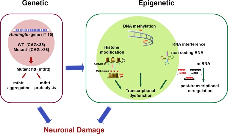
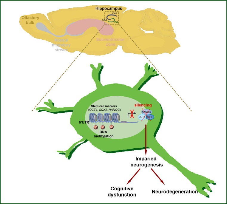
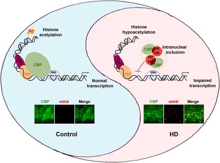
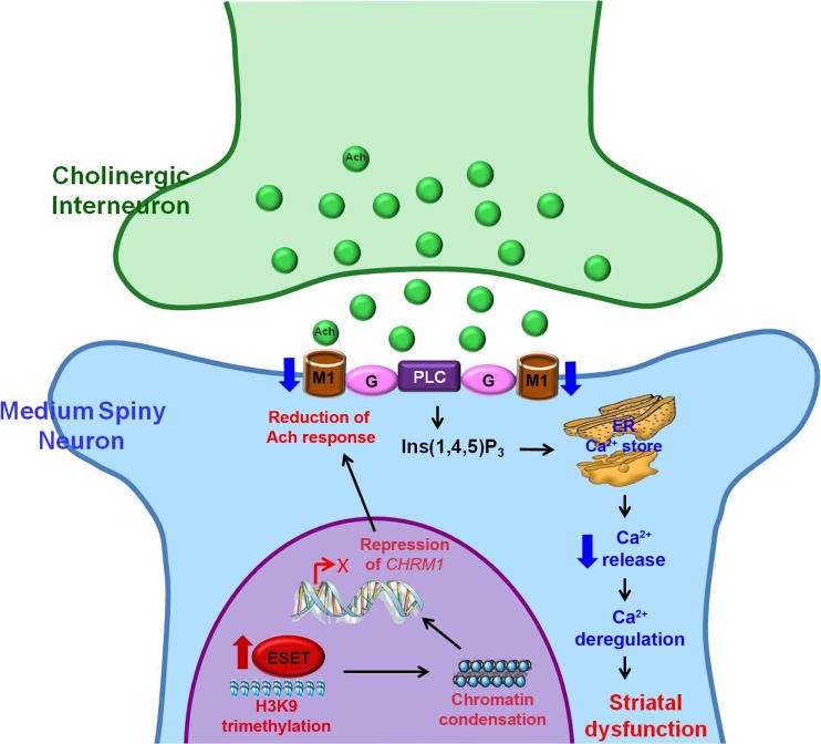
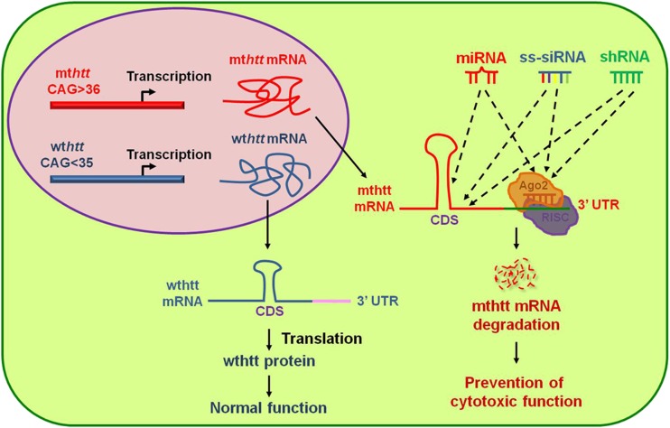
Huntington's disease (HD) is an incurable and fatal hereditary neurodegenerative disorder of mid-life onset characterized by chorea, emotional distress, and progressive cognitive decline. HD is caused by an expansion of CAG repeats coding for glutamine (Q) in exon 1 of the huntingtin gene. Recent studies suggest that epigenetic modifications may play a key role in HD pathogenesis. Alterations of the epigenetic "histone code" lead to chromatin remodeling and deregulation of neuronal gene transcription that are prominently linked to HD pathogenesis. Furthermore, specific noncoding RNAs and microRNAs are associated with neuronal damage in HD. In this review, we discuss how DNA methylation, post-translational modifications of histone, and noncoding RNA function are affected and involved in HD pathogenesis. In addition, we summarize the therapeutic effects of histone deacetylase inhibitors and DNA binding drugs on epigenetic modifications and neuropathological sequelae in HD. Our understanding of the role of these epigenetic mechanisms may lead to the identification of novel biological markers and new therapeutic targets to treat HD.


Figures

Fig. 1
Fig. 1
Alterations in epigenetic modifications are linked to the pathogenesis of Huntingdon’s disease (HD). Genetic mutation of the huntingtin (htt) gene (known as interesting transcript 15 (IT15)) leads to epigenetic alterations in neurons. DNA methylation is altered in the promoter region of neuronal genes in HD. Altered gene transcription in HD is associated with post-translational modifications of histone and abnormal nucleosomal dynamics. Neuronal gene expression is turned on (active) or off (silenced) depending on the dynamic status of histone acetylation versus methylation, respectively. Changes in noncoding RNA (ncRNA) and microRNA (miRNA) activity can deregulate gene expression at the transcriptional and post-transcriptional levels. Both aggregates and fragments of mutant htt (mthtt) may cause significant epigentic alterations that lead to synaptic and, ultimately, neuronal damage and loss in HD. The mechanisms by which these pathogenic insults trigger epigenetic modifications remain to be determined. WT = wild-type
Fig. 2
Fig. 2
DNA methylation deregulates neurogenesis in Huntingdon’s disease (HD). Regional-specific neural stem and progenitor cells turn into mature neurons of central nervous system by the process of neurogenesis. In HD, 5′-untranslated region (UTR) promoters of stem cell-related genes (octamer-binging transcription factor 4 (OCT4), SRY (sex determining region Y)-box 2 (SOX2), and Nanog homeobax (Nanog)) are methylated and neurogenesis is affected. Impaired neurogenesis results in cognitive dysfunction and could be an important epigenetic marker of neurodegeneration in HD
Fig. 3
Fig. 3
A scheme illustrates how mutant huntingtin (mthtt) contributes to cyclic adenosine monophosphate response element-binding protein (CREB) binding protein (CBP) dysfunction in Huntingdon’s disease (HD). In normal conditions, CBP maintains the acetylation status of histone through histone acetyltransferase activity and regulates the initiation of transcription by interacting with transcriptional complexes in a gene context-dependent manner. In HD, mthtt sequestrates CBP in nuclear inclusions (aggregate formation) and disrupts CBP-dependent histone modification and transcription. Consequently, imbalanced transcription and altered chromatin remodeling leads to neuronal damage resulting in cognitive dysfunction and other symptoms in HD. The confocal images show that colocalization of mthtt (red) and CBP (green) is found in nuclear inclusions of the hippocampus of HD (R6/2) mouse. TFs = transcription factors; TAF = TATA-binding protein (TBP)-associated factor; RNA Pol II = RNA polymerase II
Fig. 4
Fig. 4
A scheme represents an epigenetic mechanism that abnormal activity of histone H3K9-specific methyltransferase (ESET) leads to synaptic failure and striatal dysfunction in Huntingdon’s disease (HD). ESET-induced and H3K9me3-mediated heterochromatin condensation results in the repression of the CHRM1 gene and subsequent reduction of CHRM1 protein in medium spiny neurons (MSNs). Down-regulation of CHRM1 fails to respond to acetylcholine (Ach) from cholinergic interneurons and to transduce the G-protein-coupled intracellular Ca2+-dependent signaling pathway, which affects on the synaptic function of MSNs. Consequently, deregulation of CHRM1-dependent striatal synaptic function contributes to neurodegeneration in HD. This figure is reproduced from [69]. PLC = phospholipase C; Ins(1,4,5)P3 = inositol 1,4,5-triphosphate; ER = endoplasmic reticulum
Fig. 5
Fig. 5
Therapeutic strategies using noncoding RNAs [microRNA (miRNA), small hairpin RNA (shRNA), and single-stranded small interfering RNA (ss-siRNA)] to nullify mutant huntingtin (mthtt) expression. The mthtt gene encodes cytotoxic mutant htt protein while wild-type htt (wthtt) expresses htt protein that functions in vesicle trafficking and neuronal survival. The development of nucleic acid therapy by non-coding small RNAs is an ideal approach to selectively silence mthtt without affecting the expression of the wild-type allele. miRNAs, ss-siRNAs, and shRNAs can target either coding sequence (CDS) or the 3-untranslated region (UTR) of mthtt messenger RNA (mRNA). Consequently, they participate in mRNA degradation through Argounate (Ago) and the RNA-induced silencing complex (RISC)-dependent pathway [114, 126]

References

    1. Myers RH, MacDonald ME, Koroshetz WJ, et al. De novo expansion of a (CAG)n repeat in sporadic Huntington’s disease. Nat Genet. 1993;5:168–173. - PubMed
    1. Kremer BP, Goldberg S, Andrew J, et al. A worldwide study of the Huntington’s disease mutation the sensitivity and specificity of measuring CAG repeats. New Engl J Med. 1994;330:1401–1406. - PubMed
    1. Huntington G. On chorea. Med Surg Rep (Philadelphia) 1872;317–321.
    1. Jergelsma G. Nue anatomische befunde bei paralysis agitans und bei chronischer progressive chorea. Neurol Centralbl. 1908;27:995–996.
    1. Bruyn GW, Bots GTAM, Dom R. Huntinton’s chorea: Current neuropathological status. In: Chase TN, Wexler NS, Barbeau A, editors. Huntington’s disease. Advances in neurology. New York: Raven Press; 1979. pp. 83–93.

Publication types