. 2014 Aug;72(2):347-61.
doi: 10.1002/mrm.24924. Epub 2013 Sep 4.

Nonrigid autofocus motion correction for coronary MR angiography with a 3D cones trajectory

Affiliations

Nonrigid autofocus motion correction for coronary MR angiography with a 3D cones trajectory

R Reeve Ingle et al. Magn Reson Med. 2014 Aug.

Abstract

Purpose: To implement a nonrigid autofocus motion correction technique to improve respiratory motion correction of free-breathing whole-heart coronary magnetic resonance angiography acquisitions using an image-navigated 3D cones sequence.

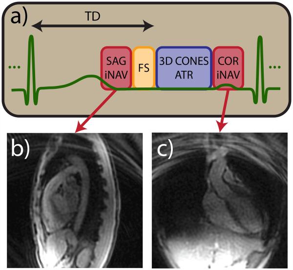
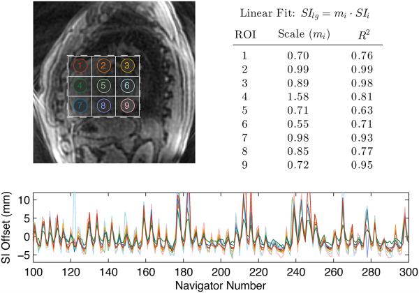
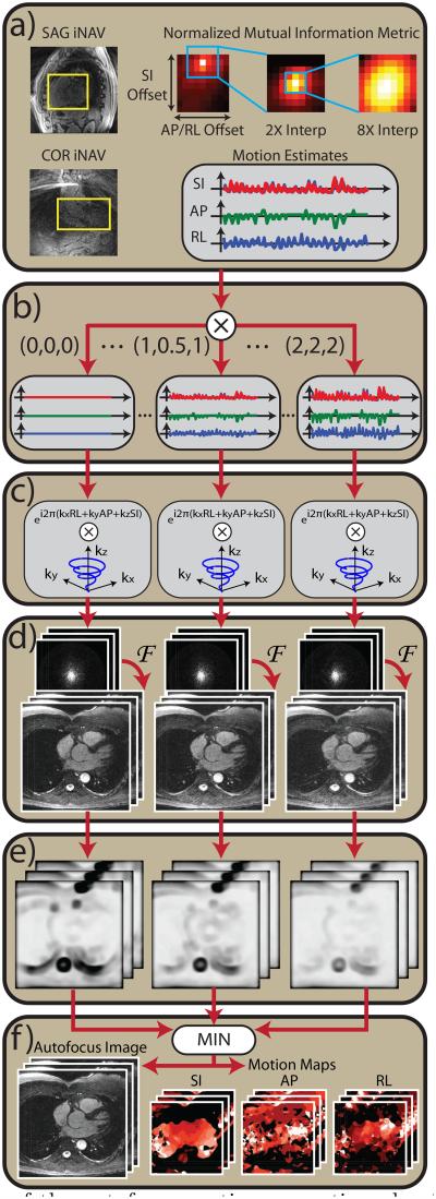
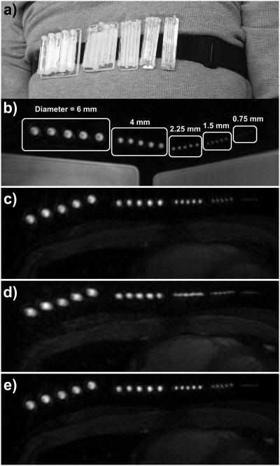
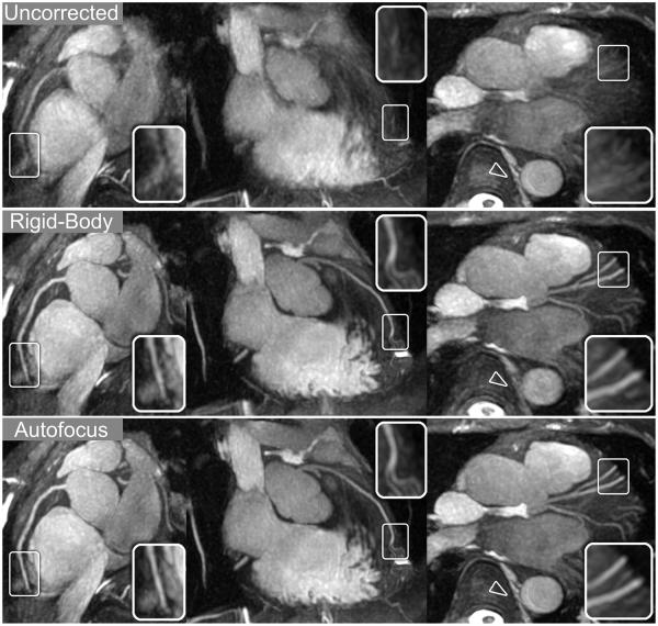
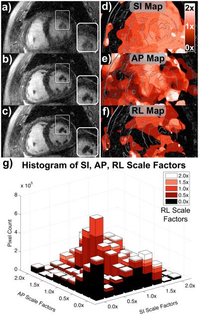
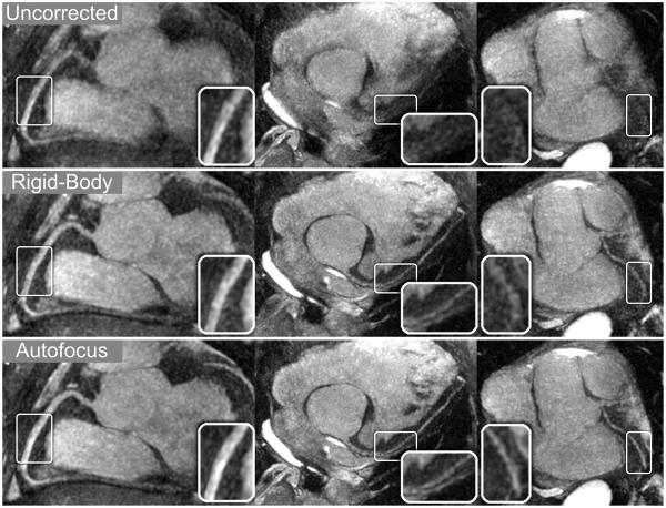
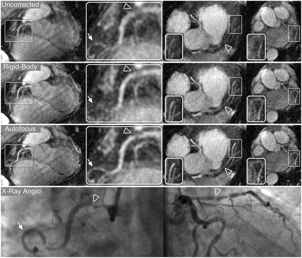
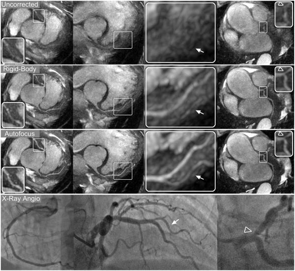
Methods: 2D image navigators acquired every heartbeat are used to measure superior-inferior, anterior-posterior, and right-left translation of the heart during a free-breathing coronary magnetic resonance angiography scan using a 3D cones readout trajectory. Various tidal respiratory motion patterns are modeled by independently scaling the three measured displacement trajectories. These scaled motion trajectories are used for 3D translational compensation of the acquired data, and a bank of motion-compensated images is reconstructed. From this bank, a gradient entropy focusing metric is used to generate a nonrigid motion-corrected image on a pixel-by-pixel basis. The performance of the autofocus motion correction technique is compared with rigid-body translational correction and no correction in phantom, volunteer, and patient studies.

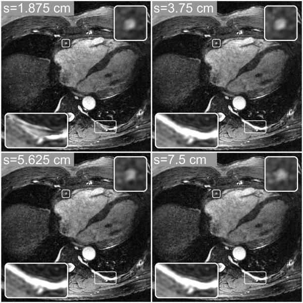
Results: Nonrigid autofocus motion correction yields improved image quality compared to rigid-body-corrected images and uncorrected images. Quantitative vessel sharpness measurements indicate superiority of the proposed technique in 14 out of 15 coronary segments from three patient and two volunteer studies.

Conclusion: The proposed technique corrects nonrigid motion artifacts in free-breathing 3D cones acquisitions, improving image quality compared to rigid-body motion correction.

Keywords: autofocus; coronary angiography; motion correction; navigator image; nonrigid.


Figures

Figure 1
Figure 1
Cardiac-triggered 3D cones acquisition scheme. (a) 3D cones data acquisition is preceded by a spiral sagittal iNAV acquisition and fat suppression module. 3D cones interleaves are acquired using an ATR-SSFP readout for additional fat suppression. Multiple imaging phases are acquired during the 3D cones data acquisition period. A spiral coronal iNAV acquisition immediately follows the cones data acquisition. Representative sagittal (b) and coronal (c) iNAVs acquired during one heartbeat of a volunteer study are shown.
Figure 2
Figure 2
Comparison of SI motion estimates derived from different regions of the heart. SI motion estimates derived from nine small ROIs (labeled in top-left image) were compared to the SI estimate derived from a large ROI covering the heart (dashed rectangle). The SI estimates were normalized by subtracting the mean, yielding the trajectories shown in the bottom plot. The resulting trajectories have a similar cyclic pattern due to respiratory motion, but the amplitudes of the motion differ for each ROI. Motion estimates from each small ROI were fit to those derived from the large ROI using a linear model. The resulting scale factors (mi) and R2 values (top right) indicate a strong linear correlation between measurements derived from the small and large ROIs.
Figure 3
Figure 3
Block diagram of the autofocus motion correction algorithm. (a) A normalized mutual information metric is used to estimate SI, AP, and RL displacements from sagittal and coronal iNAVs. (b) A set of motion basis waveforms is constructed by amplitude-scaling the measured SI, AP, and RL displacements. (c) For each set of motion basis waveforms, 3D translational correction is applied in k-space via phase modulation. (d) Motion-compensated images are reconstructed using 3D gridding followed by a 3D FFT. (e) A localized gradient entropy focusing metric is computed for each image. (f) Pixelwise minimization of the focusing metric yields the final image and SI, AP, and RL motion maps.
Figure 4
Figure 4
Autofocus reconstructions computed with four integration window sizes. Smaller window sizes (e.g., s = 1.875 and 3.75 cm), can better model localized motion variations, yielding better nonrigid motion compensation. However, rapid motion variations permitted by small window sizes (e.g., s = 1.875 cm) can yield discontinuities in the final image (bottom-left inset images), where neighboring pixels are reconstructed with very different motion scales. Larger window sizes (e.g., s = 5.625 and 7.5 cm) lead to more spatial filtering of the metric, yielding smooth and gradual motion variations. These images have fewer artifacts and image discontinuities from rapid motion variations, but they also show increased blurring of some features (top-right inset images) due to poorer nonrigid motion modeling.
Figure 5
Figure 5
Free-breathing study with resolution phantom. (a) Photograph showing the resolution phantom strapped around the chest of a volunteer. The phantom consisted of five groups of five equally spaced vials, with inner diameters of 6, 4, 2.25, 1.5, and 0.75 mm. (b) An axial slice through the phantom is shown using a static 3D cones acquisition with the phantom strapped around a large doped-water phantom. The inner diameters of each vial are labeled. (c–e) A free-breathing 3D cones acquisition was carried out with the phantom strapped around the chest of a volunteer. An axial slice through the phantom is shown from images reconstructed with (c) no motion correction, (d) rigid-body translational motion correction using SI, AP, and RL trajectories derived from iNAVs located on the volunteer’s heart, and (e) autofocus motion correction using the same iNAV measurements as (d).
Figure 6
Figure 6
Reformatted thin-plane MIPs for volunteer A. Reconstructions using no motion compensation (top row), rigid-body translational motion compensation (middle row), and autofocus motion compensation (bottom row) were reformatted to show the RCA (left column), LAD (middle column), and LCx (right column). Rigid-body correction yields significant sharpening of the coronary arteries and cardiac features while blurring non-cardiac features such as the spinal column and descending aorta (arrowheads). Autofocus motion correction yields subtle improvements in the depiction of the coronary arteries (magnified in inset images) and is able to simultaneously sharpen non-cardiac features.
Figure 7
Figure 7
Short-axis reformats for patient C. Oblique short-axis reformats were reconstructed with no motion correction (a), rigid-body translational motion correction (b), and autofocus motion correction (c). The depiction of both non-cardiac and cardiac features (e.g., papillary muscles, magnified in inset images) improves significantly with autofocus motion correction. SI motion maps (d), AP motion maps (e), and RL motion maps (f) were derived from the autofocus algorithm and reformatted into the same oblique plane as (a–c). Lighter shades of red correspond to motion scales near 2× (white), and darker shades of red correspond to motion scales near 0× (black). The chest wall has little SI and RL motion, but moderate AP motion. An outline of the image features is superimposed on the motion maps for reference. A histogram of the SI, AP, and RL scale factors selected by the algorithm is shown in (g).
Figure 8
Figure 8
Reformatted thin-plane MIPs for patient A. Reconstructions using no motion compensation (top row), rigid-body translational motion compensation (middle row), and autofocus motion compensation (bottom row) were reformatted to show the RCA (left column), LAD (middle column), and LCx (right column). Rigid-body correction yields significant sharpening of the coronary arteries, and further improvements in vessel depiction result from autofocus motion correction. The improved vessel depiction is particularly noticeable in the inset images, which show magnified segments of each vessel.
Figure 9
Figure 9
Reformatted thin-plane MIPs for patient B. Reconstructions using no motion compensation (first row), rigid-body translational motion compensation (second row), and autofocus motion compensation (third row) were reformatted to show the RCA (left columns), LAD and proximal RCA (right-center column), and LCx (right column). Nonrigid autofocus motion correction yields the best depiction of the coronary arteries. Significant improvements in vessel sharpness can be seen in distal segments of the RCA (arrows), LAD, and LCx (inset images). Stenoses in the RCA (arrowheads, magnified in left-center column) and LAD (arrowheads, right-center column) are well-depicted after autofocus motion correction and well-correlated with x-ray angiograms (bottom row).
Figure 10
Figure 10
Reformatted thin-plane MIPs for patient C. Reconstructions using no motion compensation (first row), rigid-body translational motion compensation (second row), and autofocus motion compensation (third row) were reformatted to show the RCA and LAD (left column), LAD system (center columns), and LCx (right column). Rigid-body correction yields significant sharpening of the coronary arteries, and further improvements in vessel depiction result from autofocus motion correction. The improved vessel depiction is particularly noticeable in the magnified sections of the LAD and first diagonal (arrows, right-center column), RCA, and LCx (inset images). A 50% narrowing in the proximal LAD was identified on x-ray angiograms (arrowhead, bottom row) and is well-depicted after autofocus motion correction.

References

    1. Scott AD, Keegan J, Firmin DN. Motion in cardiovascular MR imaging. Radiology. 2009;250:331–351. - PubMed
    1. Goldfarb JW, Edelman RR. Coronary arteries: breath-hold, gadolinium-enhanced, three-dimensional MR angiography. Radiology. 1998;206:830–834. - PubMed
    1. Li D, Carr JC, Shea SM, Zheng J, Deshpande VS, Wielopolski PA, Finn JP. Coronary arteries: magnetization-prepared contrast-enhanced three-dimensional volume-targeted breath-hold MR angiography. Radiology. 2001;219:270–277. - PubMed
    1. Niendorf T, Hardy CJ, Giaquinto RO, Gross P, Cline HE, Zhu Y, Kenwood G, Cohen S, Grant AK, Joshi S, Rofsky NM, Sodickson DK. Toward single breath-hold whole-heart coverage coronary MRA using highly accelerated parallel imaging with a 32-channel MR system. Magn Reson Med. 2006;56:167–176. - PubMed
    1. Wang Y, Rossman PJ, Grimm RC, Riederer SJ, Ehman RL. Navigator-echo-based real-time respiratory gating and triggering for reduction of respiration effects in three dimensional coronary MR angiography. Radiology. 1996;198:55–60. - PubMed

Publication types

MeSH terms